Monthly Archives: January 2016

What causes heart disease – part III

In most diseases it is best to start at the beginning and work forwards. This should be the case with cardiovascular disease (CVD) too. However, for complex reasons I found myself starting at the end, and working backwards. The main reason for this is that I had to start with certainty. Yet, almost everywhere I looked there was mush. For example, the epidemiology of CVD.

Now you would think that there would be agreement about how many people actually die from CVD in different countries and at different times. Not a bit of it.

A researcher:                                    ‘The French have a low rate of CVD.’

A N Other researcher:            ‘Oh well the French, they don’t agree with the normal definitions, they don’t classify CVD properly. Who knows what the true rate may be?’

True? False? A bit true? Taking another example. I have looked at the figures from the US and, you know what. Not a single person died of Ischaemic Heart Disease before 1948. Amazing. What was protecting them? [Dying from IHD is what you would also call a heart attack, or MI]. What was protecting them was the fact that IHD did not exist in the US as a disease classification, before 1948.

This then changed. In 1948 the World Health Organisation was created, and one of the first things they did was to create an International Disease Classification system (ICD). Heart disease is 1. Cancer is 2. (example for illustrative purposes only). Of course it is a bit more complex than that. Just to look in more detail at Ischaemic Heart Disease: [See box]:
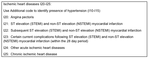
IschemicHD

Not every country took up the ICD system. Until 1968 the French did not use the ICD codes (so I am told, which no doubt means this is not true). Therefore, in France, statistics on deaths from IHD in France, before this date, are completely unreliable.

It goes without saying that, before 1948, no-one else used the ICD system either, because it did not exist. So, what can we tell about the epidemiology of CVD before 1948? Nothing. Or at least nothing you could hang your hat on. IHD would have been mixed within a much broader ‘Heart Disease’ in the death certificate statistics. And heart disease could mean almost anything, from cardiomyopathy to pericarditis, to atrial fibrillation.

Even after the ICD system was introduced, and even after France came on board, many countries clearly did not use it in the same way. Which is why the WHO set up the MONICA study.

‘The MONICA (Multinational MONItoring of trends and determinants in CArdiovascular disease) Project was established in the early 1980s in many Centres around the world to monitor trends in cardiovascular diseases, and to relate these to risk factor changes in the population over a ten year period. It was set up to explain the diverse trends in cardiovascular disease mortality which were observed from the 1970s onwards. There were total of 32 MONICA Collaborating Centres in 21 countries. The total population age 25-64 years monitored was ten million men and women. The ten year data collection was completed in the late 1990s, and the main results were published in the following years. The data are still being used for analysis.’

It was also an attempt to see if different countries were actually looking at the same diseases, and classifying them in the same way. Even after that, the data was still not absolutely clear cut, as further studies were then set up to see if the US system ARIC, and MONICA, actually matched each other. This was 1984.

‘To foster collaboration between the World Health Organization MONICA Project and the NHLBI Study of Atherosclerosis Risk in Communities (ARIC). To ensure that valid comparisons could made between findings in MONICA and ARIC by supporting activities to standardize coding, classification, and analysis of coronary and stroke events, risk factors, and medical care according to MONICA protocol.’

In simple terms, the US has its system, ARIC, and Europe had its system MONICA. Do they actually match? In short we can see that, even as late as 1984, there was clear uncertainty about how diagnoses were being made and how data were being gathered around the world. Did it all match, or not.

Given such uncertainly on both definition and diagnosis, can we say that the US epidemic of CVD in the 1960s actually happened. Or were doctors just putting IHD on death certificates when they didn’t really know what killed the patient. Personally, I think the epidemic did occur. Actually I think it happened a bit earlier. It is my belief that it took a while for US doctors to start using the new-fangled WHO ICD system.

Anyway, the point I am trying to make is that it is incredibly difficult to find the ‘bedrock.’ By which I mean facts that are inarguable. Things you can base your thinking on that are absolutely true, or that are as close to absolutely true, as possible.

Which is why I ended up at the end, the formation of the final, often fatal, blood clot. A blood clot which, generally, forms over an existing atherosclerotic plaque. There is widespread agreement that this is the case. So we can, I think safely, start here.

[There are, undoubtedly other things going on, such as sympathetic stress, mitochondrial damage and acidosis with heart muscle. that play a hugely important role. But the clot is, usually the final event

It is also widely agreed that factors which increase blood clot formation (thrombophilc factors) increase the risk of dying from CVD, and that things that reduce blood clotting reduce the rate of death from CVD. Here are a few things that increase the risk of blood clots forming, in no particular order:

  • Dehydration
  • Waking up in the morning/getting up in the morning
  • Acute physical stress
  • Acute psychological stress
  • Having a high fibrinogen level
  • Diabetes
  • Cocaine use
  • Smoking
  • Cushing’s disease

Here are some of the things that reduce the risk of blood clots

  • Haemophilia
  • Von Willibrand Disease
  • Aspirin
  • Moderate alcohol consumption
  • Clopidogrel
  • Yoga
  • Regular exercise

I suppose I should add that all of the things that increase the risk of blood clotting also increase the risk of death from CVD, and vice versa.

This is hardly a complete surprise. If blood clots kill you, things that reduce blood clots will prevent you from dying, and vice-versa. Let us not fall to the ground in stunned amazement over this statement of the bleeding obvious.

At this point, and slightly out of sequence, I would like to introduce statins to the list of factors that reduce the risk of blood clots

Readers of this blog know that I am not keen on statins, to say the least. However, if the studies are to be believed, they do reduce the risk of CVD. Not to any great extent, but the effect certainly does exist. Many people use this fact to attack my view that raised cholesterol does not cause CVD. ‘Well, what about statins,’ they bellow in delight. ‘They lower cholesterol and reduce the risk of CVD. Case proven…next’

Well, as with all drugs, statins do many other things than lower cholesterol levels. For example:

‘Recent studies have shown that statins reduce thrombosis via multiple pathways, including inhibiting platelet activation and reducing the pathologic expression of the procoagulant protein tissue factor.’1

So, as they say, there. In fact, one could quite sensibly propose that statins work pretty much the same as aspirin. They are anti-coagulants, and lowering blood cholesterol is simply a nasty and unfortunate side-effect of statins.

In reality, statins have a far more important effect on CVD (through other actions also related to clot formation) that I will get to later. I just thought I would pop that statin fact in. I even provided a reference. I have not really done much referencing in this series up to now. I believe that it is very simple to type, for example, ‘regular exercise and reduced thrombus formation’ into Google and see what you get. Or ‘Yoga and reduced blood coagulation.’

Where was I? Oh yes. Things that increase blood clot formation are more likely to kill you from CVD, and vice versa. Nothing controversial here. But the potentially controversial bit starts right here.

Are there two processes or one?

Currently, whilst conventional thinking on CVD accepts that blood clot formation is almost always the final event in CVD. This represents a completely separate process to the development of the atherosclerotic plaque itself. In short, we have two unrelated physiological processes:

  • Plaque formation
  • Clot formation on top of plaque

I apologize for saying, essentially, the same thing in different ways. But I think it is important.

Strange then, is it not, that plaque formation and clot formation share so many risk factors? Smoking, for example. Diabetes, for example. In fact, you could say (with certain provisos) that the risk factors for plaque formation and blood clot formation, are exactly the same.

Which gives one to think. Well it certainly gave me to think. Could it be that plaque formation, and blood clot formation, are simply two different manifestations of exactly the same underlying disease process. From a pure scientific perspective, I liked the idea. I liked it because it seems clumsy to have a disease, CVD, that is made up of two, essentially unelated processes

In medicine, as in all of science, one single disease process always looks much better, much cleaner, and much more likely to be right. This is the principle of Occam’s razor:

‘The principle in philosophy and science that assumptions introduced to explain a thing must not be multiplied beyond necessity, and hence the simplest of several hypotheses is always the best in accounting for unexplained facts.

Next: The four step process of CVD

References:
1: http://www.ncbi.nlm.nih.gov/pubmed/24422578

What causes heart disease – part II

[By heart disease I mean, the development of atherosclerotic plaques in large arteries. Mainly the coronary arteries (supplying blood to the heart) and carotid arteries (supplying blood to the brain). This, I will refer to as Cardiovascular Disease CVD. Not entirely accurate, but language never is.]

At this point, I am going to start at the end. What kills people? (Or what causes myocardial and cerebral infarctions). You might think that this was clear cut, but of course it is not, far from it. For example, there is a major cause of death from ischaemic strokes that has nothing whatsoever to do with CVD. This is Atrial Fibrillation.

Atrial fibrillation (AF), is a condition where the upper chambers in the heart (atria) do not contract in a regular and co-ordinated fashion. Instead, they fibrillate – AF definition : ‘(of a muscle, especially in the heart) make a quivering movement due to uncoordinated contraction of the individual fibrils.’ AF is pretty common.

People who have AF tend to develop blood clots in the atria. These can break loose, and are then ejected from the heart into other parts of the body. Quite commonly these clots travel into the brain. As the artery the clot is traveling down narrows, the clot gets stuck, and blood supply is cut off, leading to an area of ‘ischaemia’ (no oxygen) and a stroke. A cerebral infarction.

Which means that it is perfectly possible to have strokes (cerebral infarctions) that are unrelated to CVD. If, that is, you define CVD as the development of atherosclerotic plaques. You can also have strokes where an artery in the brain bursts, causing bleeding into brain tissue, which is called a haemorrhagic stroke. This is clinically indistinguishable from an ischaemic stroke. You need a brain scan to see what type of stroke has happened.

Ergo, whist death from a stroke is clearly a form of cardiovascular disease, many strokes have nothing whatsoever to do with atherosclerotic plaques – which is what I am calling CVD.

Equally you can die from something commonly defined as a ‘heart attack’ which has nothing to do with atherosclerotic plaque development either. You can, for example, develop a fatal arrhythmia. This is where the conduction system in the heart goes wonky, the heart stops contracting regularly, and you die. [Of course, quite often this happens as part of a myocardial infarction].

If we put aside these forms of dying of strokes and heart attacks, we can then focus more clearly on the event that kills you with CVD? Which is, in general, a clot forming on a vulnerable plaque, and blocking an artery, leading to a myocardial infarction (heart attack).

Looking at strokes. If a clot forms in a carotid artery, it does not tend to block the artery completely. Instead, a part of the clot breaks off, and travels up into the brain where is gets stuck – causing a stroke (as per AF).

But… there are those who disagree with this simple model. Mainly with regard to heart attacks. I am fully aware of a growing movement which states that the myocardial infarction (heart attack) happens first, then the clot forms afterwards. (Incidentally, this concept is not new; it was first proposed over eighty years ago. You may think that is seems completely mad. However, there is strong evidence that would appear to support this ‘reverse’ hypothesis’ (infarction first, then the blood clot forming in the artery).

For example, in many cases after a confirmed and accurately diagnosed myocardial infarction, you cannot find any blood clot in the artery leading to the infarcted area. In other cases, you can find a blood clot that is several days, or weeks old. This age of the clot can be established because of the ‘evolved’ state of the thrombus. In short, there is no ‘temporal’ connection between the blood clot forming and the heart attack occurring.

On the other hand, you can find an acute blockage of a coronary artery that has not caused any symptoms, let alone a myocardial infarction.

Anyway, if you try to bring these facts together you find that:

  • Myocardial infarctions can occur without any clot being found in an artery
  • A clot can form in coronary artery days, or weeks, before any symptoms of an MI
  • A clot can fully obstruct the coronary artery, without causing a myocardial infarction

Given these facts (facts which, incidentally, are not in dispute), you can make a pretty strong case that there is no causal association between a blood clot blocking a coronary artery, and an MI taking place. Instead people can, and indeed do, argue that the process is the other way around. Infarction first, then clot.

Perhaps, now, you can begin to understand why it has taken me thirty years to try and work out the underlying process of CVD. At times I have thought that there isn’t any… but that is another story, for another place

The reverse hypothesis – why it is not correct

I have studied the ‘reverse hypothesis’ – if that is a reasonable term for it – for many years, and I believe that it is wrong. The primary cause of a heart attack is simply a blood clot blocking a coronary artery. However, there are two major complications that lead to the apparent contradictions listed above. In no particular order they are the following:

  • An infarction does not mean that heart muscle dies
  • Collateral circulation develops

What is an infarction?

Cardiology is, unfortunately, dominated by highly simplistic thinking. Namely, plaque develops, clot forms, infarction occurs. The infarction occurring within minutes of the clot formation.

But this is nothing like the reality of what actually happens. Heart muscle, like all tissues in the body, is enormously complicated. If you suddenly reduce the blood supply, it can do several different things. It can infarct, it can hibernate, or it can do nothing much.

Just to look at infarction. The dictionary definition is… ‘obstruction of the blood supply to an organ or region of tissue, typically by a thrombus or embolus, causing local death of the tissue.’ Well, I’ve got news for you, heart muscle does not die (not unless the organism surrounding it dies).

Infarction, in the case of myocardial infarction, does not mean death of the tissue. Instead, the heart muscle undergoes a complex transformation into a different cell type. One that needs far less oxygen to survive and one that cannot do the contracting thing. But it is not dead. Because dead cells become necrotic and necrotic cells disintegrate. After a heart attack do you see disintegrated areas of the heart? No, you most certainly do not. You see a form of scar tissue developing.

There is also a halfway house that lies between infarction, and nothing happening. This is ‘hibernation’, a state whereby heart muscle simply decides to stop contracting/beating (to save oxygen use). If you do MRI scans on the heart, in those with known heart disease, such ‘silent’ regions are a relatively common finding. Sometimes these regions wake up and start beating again, sometimes they do not. Sometimes they go on to infarct.

Adding further to the complication, if a coronary artery starts to narrow, the heart will create small ‘collateral’ blood vessels to get round the narrowing, and keep the blood supply up. When, and if, the artery fully blocks, the collateral circulation will maintain the blood flow, and so nothing very much will happen, even if the coronary artery is fully blocked. Many people survive on collateral circulation alone.

All of which means that, after an artery blocks, one of three things can happen:

  • There is a sudden infarction (could be large enough to be fatal)
  • The heart muscle decides to hibernate, if there is sufficient collateral circulation to keep things ticking along
  • Nothing much happens. If there is high level of collateral circulation, the heart just carries on much as before.

Only in the first case will the obstructive blood clot closely precede the heart attack. In case two the hibernating heart muscle may later decide to infarct, if the circulation does not improve. This can happen under periods of high physical or psychological stress. Thus the formation of the blood clot can precede the infarction by days, weeks or months. Indeed, the clot may have been fully cleared away by the time infarction occurs.

In short, the apparent contradictions to the: clot → blockage → infarct hypothesis can be explained, reasonably easily. So long as you realise that what happens inside the heart is not a case of simple pipe-work, where there a fixed number of tubes (arteries) supply oxygen to a pump (the heart). If you block one tube, the pump is immediately damaged.

Heart attacks and strokes

However, my main reason for disbelieving the ‘reverse hypothesis’ is that the standard model works for both heart attacks and (most) ischaemic strokes.

In ischaemic strokes, as mentioned before, a blood clot forms over a plaque in a carotid artery. This rarely blocks the artery; as carotid arteries are much wider bore than coronary arteries. However, what happens next – after a variable time period – is that the clot breaks off and travels into the brain.

This is, essentially, exactly the same process as a heart attack – except the clot lodges further down the vascular tree. Not only are the processes of stroke and heart attack virtually identical, the risk factors for both are virtually identical. Perhaps most telling is the fact that people with plaques in their coronary arteries almost always have plaque in their carotid arteries, and vice-versa. For example, here is the title of a paper on this topc: ‘Tight relations between coronary calcification and atherosclerotic lesions in the carotid artery in chronic dialysis patients.’1

This is further supported by a study that has just come out, and published in BMJ open. Key points were:

  • We studied the risk of heart disease following a stroke in those patients with no cardiac history. This study is the largest of its kind and, by bringing together multiple data sets, robustly quantifies the risk of heart disease following stroke. As with all meta-analyses, the main limitation of this work relates to publication bias.
  • Most patients with stroke die of heart disease.
  • One in three patients with ischaemic stroke with no cardiac history have more than 50% coronary stenosis.
  • 3% are at risk of developing myocardial infarction within a year of their stroke.
  • Patients with stroke need to be screened for silent heart disease and appropriate and aggressive management of total cardiovascular risk factors is required2.

In short, the two conditions are the same. It is beyond any reasonable doubt that with ischaemic stroke, and myocardial infarctions, we are looking at the same underlying disease. To be frank I don’t think may people would disagree with this. But it does lead to a critical point. Namely has anyone, ever, argued that cerebral infarctions (strokes) happen before the blood clot blocks an artery in the brain?

No they have not. Because to do so would, quite frankly, be bonkers. We can be absolutely certain that blood clots cause infarctions in the brain. Yet many people quite strongly argue that with myocardial infarction it is the other way around. For the reasons outlined above I do not, and cannot, believe this. There is only one process, and no need to start searching for another.

You may wonder why I have gone off in such a wide detour here? There are two reasons. First to make it clear that this area is gigantically complex. Secondly, to reinforce the point that wherever and however you look at CVD, alternative hypotheses have been proposed. Until you have tracked them down, and examined them fully, you cannot really move on.

Next: Some answers.

References:
1: http://www.ncbi.nlm.nih.gov/pubmed/20470277

2: http://bmjopen.bmj.com/content/6/1/e009535.full

What causes heart disease?

I have been somewhat silent on this blog for a while. Mainly because I have been putting together ten thousand words on the true cause of heart disease. Of course, by heart disease I mean the thickenings and narrowings in the larger arteries in the body (atherosclerotic plaques). I am also focussing almost entirely on the arteries supplying blood to the heart (coronary arteries), and the main arteries that supply blood to the brain (carotid arteries).

Whilst atherosclerotic plaques can develop in other arteries that supply, for example, the kidneys, or the bowels, problems here are generally less common, and less severe – although not always. In general, however, the main killers in ‘heart disease’ are heart attacks and strokes (not all strokes, only the most common form of stroke, an ischaemic stroke). So, at the risk of becoming over-pedantic, and simultaneously sloppily inaccurate, I am calling heart disease Cardiovascular Disease (CVD), and looking at heart attacks and strokes.

With that out of the way, what causes cardiovascular disease (CVD)? Whilst it took me only a few days of research, many years ago, to realise that the diet-heart/cholesterol hypothesis was clearly nonsense. It has taken over thirty years to work out what is actually going on. In truth I could not truly progress until it suddenly dawned on me that I should not be looking for causes. For that is a mugs game.

Once you start looking for causes, you find that there is almost nothing that a human can ingest, or do, that has not been claimed to be a cause of CVD, or a cure for CVD. In many cases both… simultaneously. In 1981 a paper was published in the journal Atherosclerosis which outlined several hundred possible ‘factors’ involved in causing or preventing CVD. Today, if you hit Google, or Pubmed, I can guarantee that you could find several thousand different factors. If, that is, you could be bothered.

If you could be bothered, what could you possibly make of ten thousand different things involved in CVD in one way or another? Can they all be true risk factors? Some of them are certainly only associations, not causes. A few are simply statistical aberrations, found in one study and contradicted in another. Even removing them, there are so many, so very very many. Pick your favourite and trumpet it to the world. Vitamin K, Vitamin D, coffee, leafy green vegetables, omega 3 fatty acids, intermediate chain monounsaturated fats, HDL raising agents, selenium, lowering homocysteine…. on and on it goes.

Is there any other hypothesis where you have to fit in ten thousand different factors? No, there is not. Yet no one has been put off identifying more and more things. This is why, I believe, we have such a terrible mess. I amuse myself sometimes looking at the knots the cholesterol hypothesis ties itself into to.

Just to give one example. We have had ‘good’ cholesterol and ‘bad’ cholesterol for some time now. More recently we have ‘bad good’ cholesterol (raised HDL increasing CVD risk during the menopause), and ‘good bad’ cholesterol (light and fluffy LDL that protects against CVD). Now, that’s what I call a non-disprovable hypothesis. A risk factor that can be good, or bad. Or ‘Good bad’ and ‘bad good’.

Whilst contemplating such nonsense it came to me, in a moment of the blindingly obvious, that in order to understand CVD I had to move away from trying to fit ten thousand factors into the biggest intellectual jigsaw known to man, and move on. I had to know what the actual process is. What is actually happening in the arteries.

Once you start to look at CVD through this window, you suddenly realise that very little is ever written on this topic. The world famous cardiology bible ‘Braunwald’s Heart Disease’ is virtually silent on the matter. Or at least it was when I looked through it a few years ago.

In a massive book on heart disease, the process of CVD development is covered in less than half a page. The cholesterol hypothesis itself is usually left completely unexplained. Or there are gaping holes, and bits that you just have to take on faith. Raised LDL leaks/travels/gets into the artery wall where it creates inflammation and plaques develop. The end.

Then you start to ask, so why do plaques never develop in veins? Same structure as arteries, same level of LDL. Why do plaques never develop in the blood vessels in the lungs (pulmonary arteries and veins?). What has oxidised LDL got to do with it? Where does oxidation occur? How does LDL leak into coronary arteries, and carotid arteries, but cannot leak into arteries within the brain itself?

Questions, questions, questions and almost no decent answers. There is a kind of collective brouhaha noise with a lot of ‘well it just does’ thrown in when you start to ask. ‘Explain again, how does LDL get into the arterial wall. Each step please?’. You are usually met with perfect anti-Popparin logic. We know that raised cholesterol causes heart disease, so it must get into the arterial wall using some mechanism or other. And look, there is cholesterol in atherosclerotic plaques. So it must get through.

Of course, it is true you can find cholesterol in atherosclerotic plaques. No-one is going to deny that. But you can also find, for example, red blood cells (RBCs). Now, you might be able to explain how LDL can pass through endothelial cells (the cells that line the arteries) in some fashion. Although I would argue that, if so, why does LDL not pass through endothelial cells in veins. And why cannot it pass through, or between, endothelial cells in the arteries with the brain?

LDL molecules, after all, are minute in comparison to an endothelial cell. However, RBCs and endothelial cells are pretty much the same size. So, please try to explain to me how a RBC finds itself within the artery wall, underneath the endothelium? Try getting one cell, virtually the same size as another, to pass through it. A very clever trick indeed.

Then, if you start exploring further, you find that the cholesterol you find in atherosclerotic plaques almost certainly comes from the cholesterol rich membranes of RBCs.

The view that apoptotic macrophages (dead macrophages) are the predominant source of cholesterol in progressive (atherosclerotic) lesions is being challenged as new lines of evidence suggest erythrocyte membranes contribute to a significant amount of free cholesterol in plaques.’1

Oh look, it seems that the cholesterol does not come from LDL. Anyway, I am jumping ahead of myself here, and getting dragged back into explaining why the cholesterol hypothesis is nonsense. Which is playing the game on the opponents’ pitch, under their rules.

The simple fact is that, to replace the Cholesterol hypothesis, there is a need to come up with something better, which actually fits all the facts. That, of course, is rather trickier as – boy – there are a lot of facts. Also, some of them may seem utterly disconnected.

My simple credo is that, if your hypothesis cannot explain everything about CVD you cannot explain anything. Attempting to do otherwise means that you are left suggesting that there are many different causes, and many different processes, all of which end up causing CVD through non-connected mechanisms. Well if that is true, then we just have to give up. Smoking causes CVD like this, LDL causes it like that, diabetes in a completely different way.

This is why I get so frustrated when people simply shrug their shoulders in a debate on CVD, and retreat to the position of inarguable logic when they tell me that CVD is ‘mutifactorial.’ To which I agree that of course it is bleeding mutlfactorial (as are all diseases). But that the statement itself is meaningless, unless you can then tell me how all the ‘multi’ factors fit together within a single, unified process.

In short, with CVD, if you are going to explain it, you need to be able to explain how, for example, the following factors increase risk, and through what single mechanism, or process. [This is not an exhaustive list by any means, but these are all definite, and potent, causes]:

  • Rheumatoid arthritis
  • Steroid use
  • Systemic Lupus Erythematosus
  • Smoking
  • Kawasaki’s disease
  • Use of Non-steroidal anti-inflammatory drugs e.g. ibuprofen, naproxen and suchlike.
  • Being a deep coal miner – especially in Russia
  • Using cocaine
  • Getting older
  • Getting up in the morning – especially on Mondays
  • Type II diabetes
  • Raised fibrinogen level
  • Cushing’s disease
  • Air pollution
  • Acute physical or psychological stress
  • Chronic kidney disease
  • Avastin – a cancer drug

Looking at one of these risk factors, System Lupus Erythematosus. Young women with this condition have, in some studies, an increased risk of CVD of 5,500%. Compare that with, for example, raised LDL. Even if you believe that it raises the risk of CVD, which is debatable, the increase in risk (as defined by mainstream research) is 66% for a 3mmol/l increase in the LDL level2. Changing the LDL level by this much takes you from low risk, to Familial Hypercholesterolaemia (FH).

If we accept that the 66% figure is, indeed, correct, we can see that SLE increases the risk 83 times more than having a very high LDL level. Or, to frame this differently. SLE increases the risk of CVD 8,300% more. Clearly, therefore, SLE has far more to tell us about what really causes CVD than raised LDL ever could. Deep coal miners in Russia have their final, fatal, heart attack aged 42, on average. Children with Kawasaki’s disease can die of a heart attack aged 3.

Here, therefore, are the real causes of CVD. Super accelerated CVD with death at a young age. No need for statistical games. This is the where the answers truly lie. Now comes the difficult bit. How can you fit them all together within a single disease process, without finding anything contradictory?

Ladies and gentlemen, it took me thirty years.

References
1: https://www.researchgate.net/publication/5958670_Free_cholesterol_in_atherosclerotic_plaques_Where_does_it_come_from

2: http://www.jbs3risk.com/pages/impact_intervention.htm