Category Archives: Cardiovascular Disease

A new paper by me – please share widely

26th August 2022

And so, after a great deal of faffing about, my article on cardiovascular disease ‘Assessing cardiovascular disease: looking beyond cholesterol’ has been made free to view.

Writing an article for a medical journal is not that difficult. Trying to submit it through Kafkaesque editorial systems, now, that is tricky. They seem incapable of understanding that I am not funded by anyone. Shock, horror, I do it simply for the love of science – or something of the sort.

As for allowing the article to be open access … don’t go there. I would rather fill in a US tax form, in triplicate and, yes, I have seen US tax forms in all their incomprehensible glory. I also spent considerable time trying to explain to the editorial team that the two risk calculators I discussed in the paper could not be referenced in the approved Vancouver style.

Vancouver style: required elements:

Author. Title [Type of medium]. Place of publication: Publisher; Date of publication [Date of update/revision; Date of citation]. Availability.

I could not use Vancouver style because there was no author, place or date of publication – to start with. They were both on-line tools used to assess cardiovascular risk. Helloooo… ever heard of the Internet. Thud!

Anyway, I was invited to write the article by Dr Eric Westman, who was the guest editor for this edition of ‘Current Opinion in Endocrinology, Diabetes and Obesity.’ In truth, I get about fifty invites a day to write articles. This is not a boast, anyone who has written almost anything that has been published in a medical journal is bombarded with such requests. New journals spring up like desert flowers after the rain.

Most of the requests are, essentially, vanity publishing. You spend ages putting together a paper that you then must pay to get published – you certainly have to pay a lot to allow open access. Usually thousands of dollars. The publisher meanwhile gains copyright. Then hardly anyone ever reads it. But, hey, you can send a copy to your mum – who will be very proud. If none the wiser what you are trying to say.

Thus, I do not respond to such requests normally. But in this case, I did. Eric Westman is a staunch ally in the crusade to look at different causal models of cardiovascular disease. Models not based on LDL/cholesterol levels.

For this edition he also invited others e.g., David Diamond to write other articles casting doubt on the LDL/cholesterol hypothesis – in the proper scientific manner. Dr Westman then paid to make them open access. A cost running into many thousands of dollars. Good man.

Anyway, here it is. https://journals.lww.com/co-endocrinology/Fulltext/9900/Assessing_cardiovascular_disease__looking_beyond.21.aspx

For those who have read my blog assiduously, or have read ‘The Clot Thickens’, none of this is new, or any surprise. However, I hope that it does add some more scientific credibility.  Here is the abstract.

Abstract

Purpose of review

The low-density lipoprotein (LDL)-cholesterol level is a weak predictor of developing cardiovascular (CV) disease and can only explain a small proportion of CV risk. It is not used to determine CV risk on either the atherosclerotic cardiovascular disease (ASCVD) calculator in the United States, or the Qrisk3 in the UK.

A study in JAMA in 2022 suggested that ‘the absolute benefits of statins are modest and may not be strongly mediated through the degree of LDL reduction’. Perhaps it is time to look beyond cholesterol to a different causal model – the ‘thrombogenic’ model of ASCVD.

Recent findings

The severe acute respiratory syndrome coronavirus 2 (SARS-CoV2) pandemic demonstrated that infectious agents damage the endothelium and the glycocalyx – the glycoprotein layer protecting underlying endothelial cells. There are numerous other conditions leading to this kind of damage, which can trigger thrombus formation, causing strokes and myocardial infarctions.

Although these are acute events, they highlight a mechanism for the development of ASCVD which centres on endothelial damage and thrombus formation as both the primary causal mechanism for acute events, and the driver behind progression towards atherosclerotic plaque development.

Summary

The cholesterol hypothesis, that a raised LDL is directly causal for ASCVD, does not adequately explain cardiovascular risk in individuals, or populations. An alternative ‘thrombogenic’ hypothesis is proposed as a more valid causal model.

Here are the key points

KEY POINTS

  • Low-density lipoprotein (LDL)-cholesterol is a weak predictor of cardiovascular risk
  • Factors that drive endothelial damage and thrombus formation greatly increase atherosclerotic cardiovascular disease (ASCVD) risk.
  • The thrombogenic can explain a number of causal risk factors that do not fit within the LDL-cholesterol hypothesis, including type II diabetes, smoking and systemic lupus erythematosus.
  • The thrombogenic hypothesis, that endothelial damage and subsequent clot formation underlies the formation and growth of plaques, may represent a better model for ASCVD.
  • There is a need to research the thrombogenic hypothesis in more depth.

Stripping away the scientific obfuscation which is now required of all scientific writing. If anyone understands what you are trying to say, you lose. The two points that I was trying to make were the following:

  • The LDL/cholesterol hypothesis is most likely wrong. It is a weak predictor of risk, at best, and cannot explain how many factors known to increase the risk of cardiovascular disease, actually cause cardiovascular disease.
  • There is an alternative hypothesis, the ‘thrombogenic’ hypothesis which can explain how, and why, many different, apparently unconnected factors actually do cause cardiovascular disease.

I hope that the readers of this blog can make as much noise about it as possible and share it as widely as possible. You may even want to read the entire paper. It is not very long, and it is not too technical. At least I don’t think so.

Thank you.

Pfizer and me – Best Buddies

21st March 2022


Someone once said to me that I really must despise the pharmaceutical industry. There are certainly times when this is true, and my anger with them is sharp … and hot.

But yet, and yet, I know many people who work in the industry, and they all seem nice, concerned about the world, caring. Trying to do good. The industry itself has also produced some great innovations and medications. Without which the world would be a much scarier and more unpleasant place.

In truth, I find the industry is a bit like capitalism. Both fantastic and dreadful. Which is a bit like humanity itself. Both fantastic and dreadful. Capable of the most amazing things, yet the darker side can be very dark indeed. Dr Jekyll and Mr Hyde.

To be frank, my personal problems with the pharmaceutical industry have mainly centred around cholesterol lowering. Various companies have made billions, nay tens of billions, nay hundreds of billions, pushing LDL-Cholesterol reduction with all their might.

However, I have oft sat with my head in my hands in despair at such nonsense. Pfizer with Lipitor (atorvastatin) pushed the hardest and made the most … and horribly distorted the entire world of cardiovascular disease research in so doing.

However, thirty years ago, and purely by chance, the planets Pfizer and Kendrick were in a strange alignment. Briefly, I found myself on the same side as Pfizer when it came to cardiovascular disease.

How could this possibly be so? Well, gentle reader, let me take you back to a time when Pfizer had a very different drug to promote, called Doxazocin. It was a type of blood pressure lowering agent known as an Alpha-1 blocker. It was not selling terribly well, so they were looking for other angles in an attempt to boost sales.

I should mention that this was before Pfizer had a statin. A time that you could refer to as the ‘BS’ era. (Or maybe we are now in the true BS era – discuss). Then, in the year 2000, Pfizer took over Warner Lambert, which just happened to have a statin called atorvastatin (Lipitor). Yes, Pfizer didn’t actually develop atorvastatin. They just bought out the company that did. Nifty move.

Anyway, in 1992, Pfizer was not much interested in lowering LDL-Cholesterol, as they didn’t have a statin. Which meant they had other fish to fry with their cardiovascular drugs. They were more focussed on blood pressure lowering. But their Alpha-1 blocker for this, doxazocin (Cardura) was a bit rubbish.

So, what to do? What to do indeed. What they did was to use the tried and trusted mechanisms of looking at different effects that this drug might have.

To explain, almost all drugs when they are launched are marketed for one indication, and that indication only – the thing that will make the most money. Achieving marketing approval, for any indication e.g., blood pressure lowering, costs vast sums of money. Which means that companies must keep their focus tight, to drive the drug through the clinical trials process.

However, almost all drugs will do many other things, and you cannot run separate clinical trials on them all. To long, too complex, too costly.

There may be some ‘magic bullet’ drug that hits one target, and one alone, and spares everything else. Sniper fire. However, with most of them, you select automatic, raise the gun over your head from behind a sheltering wall, close your eyes, and spray bullets vaguely towards the target. Hoping you manage to hit the thing you’re aiming at.

Just to give a few well-known examples from history. Aspirin was initially found to reduce pain, inflammation and temperature. It was later discovered it helps to prevent platelets (small blood cells) sticking together. So, it was also an anti-coagulant – it reduced the risk of blood clots forming – and thus reduced the risk of dying of a heart attack. Now, it is being used to prevent cancer.

Sildenafil (Viagra) was initially developed as a drug for angina. The rest, as they say is history. It ended up being marketed for erectile dysfunction because it had an unknown, at the time, ‘off-target effect’ that had nothing, directly to do with angina at all. Or, at least, no-one could see the connection at the time.

Thalidomide was originally developed as a sedative. It was then sold for treating colds, flu, nausea and morning sickness. As everyone now knows, it caused horrible deformities in the unborn child.

Coincidentally, it was discovered the very same mechanism that led to the terrible deformities, also meant it was pretty good at treating a whole series of different diseases, such as multiple myeloma and leprosy. It is now prescribed as Thalomid – it has many different names. But certainly not thalidomide.

Last time I bothered counting, statins had thirty-four off-target effects. These are sometimes called ‘pleiotropic’ effects. I suppose this makes them sound more scientific. I am only surprised that statins have not yet been repurposed as anti COVID19 drugs. Probably because they are all off-patent, thus cannot make vast sums of money.

In an attempt to pull all these strands together, if your drug is not selling that well, have a closer look at all the other things it may do. The off-target effects, the ‘pleiotropics’. It has already reached market, at great cost, and whilst you are not allowed advertise it for ‘off-label’ benefits i.e., benefits not studied in the clinical trials, you can hold educational meetings and produce educational booklets to promote these additional things.

You can, effectively, launch it again as ‘Cardura two point one’. Ladies and Gentlemen …. drum roll. ‘A new era in cardiovascular disease prevention has begun.’ Dun, dun, duuuuuuuuun!’

At which point you can purchase a few off the shelf cardiology opinion leaders to push Cardura two point one with great enthusiasm. Throw a few million – carefully labelled – free pens, sticky pads, and BP cuffs around as an appreciative gift to doctors. Take them to a free lunch, sorry, educational meeting. Bore them for ten minutes, then reward them with a prawn sandwich. Feed the sea-lions.

And lo it came to pass that Pfizer scrutinised Cardura in greater detail, and found that it, like aspirin, helped to stop platelets sticking together. Platelets are small cells that float around in the bloodstream and play the key role in blood clotting. They have been described as the conductors of the clotting orchestra.

If you stop platelets sticking together, you can reduce the risk of cardiovascular disease, as was discovered with aspirin. Aha! So, not only does Cardura lower blood pressure, but it also has ‘anti-coagulant’ properties. Even better, it reduced fibrinogen, and PAI-1 and increased TPa activity. These are all ‘good’ in reducing the formation and increasing the breakdown of blood clots. As Pfizer stated:

‘These recent studies suggest that doxazosin may have a range of significant antithrombotic effects in many patients…’

Unfortunately, in 1992 doctors had already been under heavy and continuous bombardment with the message that lowering cholesterol and blood pressure were, by far, the most important things you could possibly do to prevent cardiovascular disease.

Blood clotting? Well, aspirin was used, a bit. But this was mainly to help with the final event. The big blood clot that blocked a major artery. No-one was suggesting that blood clotting had anything to do with atherosclerosis itself.

After all, how can blood clotting possibly cause atherosclerotic plaques to develop? As everyone had already been told, and told, and told … and told, and then told some more, atherosclerotic plaques (the entities that gradually grow and narrow down arteries) were full of cholesterol. Not the remnants of blood clots.

Love and marriage may go together like a horse and carriage, but platelet aggregation and atherosclerotic plaques …. You certainly cannot get a rhyme out of that – go on, I challenge you. Ergo, it must be wrong.

It was certainly a big mountain to climb. ‘You know that stuff about cholesterol…. Sorry. Turns out we should have been looking at Platelets and blood clotting instead.’ Here from a booklet on Cardura in 1992:

Platelets and atherosclerosis progression

Several features of mature plaques, such as their multi-layered pattern, suggest that platelet aggregation and thrombus formation are key elements in the progression of atherosclerosis. Platelets are also known to provide a rich source of growth factors, with can stimulate plaque development.

Given the insidious nature of atherosclerosis, it is vital to consider the role of platelets and thrombosis in this process, and the serious events that may be triggered once plaques are already present.

Goodness me. Who said all this? Why, of course it was Pfizer … Pfizer, Pfizer, Pfizer.

However, they didn’t get very far with this story. Merck were hammering away with simvastatin (Zocor) and Bristol Myers Squibb were also promoting pravastatin (Pravachol) with relentless fervour. The world of cardiovascular disease prevention was moving even more firmly in the direction of cholesterol lowering and statins.

As King Cnut (Canute to you and me) once demonstrated, once the tide starts to come in, not even a king can stop it. And the statin wave was very powerful. On the basis that, if you can’t beat them, join them, Pfizer decided to ride that wave. So, they went out and bought a large part of it, then bought the surf-boards and the tinnies.

To their (money making) credit, they then rode the cholesterol wave exceptionally well. To be frank you would buy a used car from Pfizer. I don’t know how they got so brilliant at spraying bull-shit with yellow paint, to make it look like one-hundred per-cent gold bullion, but there is no doubt they are the masters of pharmaceutical marketing.

At which point the Kendrick and Pfizer, planets were no longer in alignment. We span off on different orbits. I was still slowly plodding along the ‘platelets and atherosclerosis progression’ path, trying to make sense of it all.

Pfizer, to flip my analogies, firmly jumped on the ‘all singing, all dancing’ cholesterol lowering bandwagon.’ Complete with dancing girls in fancy costumes, a brass band, champagne, hundreds of balloons – and all the key opinion leaders in cardiology singing ‘We’re in the money.’

We’re in the money

We’re in the money

We’ve got a lot of what it takes to get along

We’re in the money

The sky is sunny

Old man depression, you are through you done us wrong, oh

We never see a headline

‘Bout a breadline today

And when we see the landlord

We can look that guy right in the eye

Oh, We’re in the money

Come, on my honey

Let’s lend it, spend it, send it rolling around…

And lo it came to pass that Pfizer and I were no longer friends. But there was a time when I like to think we could have danced all night, and still have begged for more. I could have spread my wings, and done a thousand things, I’ve never done before. Yes, my bandwagon would have been much better. Better music too. John Martyn, Fleetwood Mac, Randy Newman, The Pretenders, Jools Holland ….

Yes, it’s a little bit funny that Pfizer knew, thirty years ago, that blood clotting and atherosclerosis were intimately connected. They had seen the research. They knew:

‘Important evidence is now emerging that the selective alpha-1 inhibitor, doxazosin, in addition to its beneficial effects on elevated blood pressure and the serum lipid profile, may help to intervene in the evolution of thrombosis, a key component of atherosclerosis.’

In my recent book The Clot Thickens, I said I would put up the Pfizer booklet for all to see. It proved a bit more of a technical challenge than I thought, but here it is. And when people tell me that atherosclerotic cardiovascular disease (ASCVD) is all due to raised cholesterol I can say, not even Pfizer believes that. Not really. Not deep in their hearts …. They have a ghost in the machine that still stalks the corridors of Pfizer HQ. And if it doesn’t, it should.

They knew, oh yes once upon a time, they knew. It’s just not very profitable for them to admit it. To quote John Martyn:

Half the lies I tell you are not true.’

Introduction

To date, most of our attempts to prevent atherosclerosis have centered on the control of hypertension and hyperlipidaemia, as well as lifestyle factors . However, recent insights into the pathology of coronary heart disease have sharpened our focus on the natural history of atheroma and its relentless progression to acute cardiac events.

Platelets and atherosclerosis progression

Several features of mature plaques, such as their multi-layered pattern, suggest that platelet aggregation and thrombus formation are key elements in the progression of atherosclerosis. Platelets are also known to provide a rich source of growth factors which can stimulate plaque development.

Given the insidious nature of atherosclerosis, it is vital to consider the role of platelets and thrombosis in this process, and the serious events that may be triggered once plaques are already present.

Fibrinolysis

If fibrinolysis is incomplete, thrombus may become incorporated into the plaque, and may cause severe stenosis. Alternatively, any residual thrombus can act as a powerful stimulant to further platelet aggregation. The growing mass of fibrin, platelets and enmeshed red cells may become solid enough to cause complete vessel obstruction.

It is significant to note that complete obstruction and myocardial infarction often develops from mild lesion initially causing less than 50% stenosis. Rupture of these plaques and the cascade of events that leads to thrombosis can occur rapidly and it now recognized as a common and major precipitant of unstable angina, myocardial infarction and sudden cardiac death. Studies suggest that thrombotic events may account for up to 90% of acute myocardial infarction.

Triggering factors in thrombosis

Hypertensive patients are known to have greater platelet adhesiveness and aggregability, which could increase clot formation at the site of plaque injury. In addition, thrombolysis is often defective in hypertension and hyperlipidaemia, which may result in an impaired ability to dissolved clots in the presence of atherosclerosis.

Clot propagation

Any residual thrombus can act as a powerful stimulant to further platelet aggregation. The growing mass of fibbing, platelets and enmeshed red-cells may become solid enough to cause complete vessel obstruction.

It is significant to not that complete obstruction and myocardial infarction often develops form mild lesions initially causing less than 50% stenosis. Rupture of these plaques and the cascade of events that leads to thrombosis can occur rapidly and it now recognized as a common and major precipitant of unstable angina, myocardial infarction and sudden cardiac deaths. Studies suggest that thrombotic events may account for up to 90% of acute myocardial infarctions.

The atherosclerotic process begins with infiltration of low-density lipoproteins or LDL into the arterial intima to create lipid-rich foam cells which form the basis of the ‘fatty streak.’ This early lesion contains large amounts of cholesterol, but its development to atherosclerosis is not inevitable. Progression appears to depend critically upon endothelial injury, caused by oxidation of LDL by the shearing forces of hypertension and by smoking.

Conclusion

With each decade, new insight is gained into how drug therapy can reduce the risk of coronary heart disease and stroke, the primary goal in the management of the hypertensive patient.
Important evidence is now emerging that the selective alpha-1 inhibitor, doxazosin, in addition to its beneficial effects on elevated blood pressure and the serum lipid profile, may help to intervene in the evolution of thrombosis, a key component of atherosclerosis.

The Document is called Pathological Triggers ‘New Insights into Cardiovascular Risk.’

Produced by Medi Cine Inc
488 Madison Avenue
New York
NY 10022

For Pfizer Inc
New York
NY 10017 Copyright 1992 Pfizer Inc. All rights reserved.

The enduring mystery of heart disease – The Clot Thickens

4th November 2021

And so, after only thirty short years of research, my book on heart disease is finally finished, and is published today. Up to now my books, and blogs, have mainly consisted of telling people what does not cause heart disease. Or to be slightly more accurate, cardiovascular disease.

Hint, it is not cholesterol, or LDL, or LDL/cholesterol, or the good cholesterol/bad cholesterol ratio – or whichever term is in favour today.

So, what does cause cardiovascular disease? Those few, those happy few, who have read this blog over the years may already know a great deal of what is in the book. For others this will be a new and, hopefully, fascinating foray into a completely different way of thinking.

The first point I want to make, then emphasize, then re-emphasize, is that there is no ‘the’ cause of cardiovascular disease. By which I mean that there is not, and never has been, any one single factor that can be considered to be ‘the’ cause. Instead, there are many. They can work by themselves or combine with others.

So you can say that, for example, diabetes is ‘a’ cause of cardiovascular disease. But you cannot say that it is ‘the’ cause. Yes ‘a’ cause but not ‘the’ cause. This is not purely semantics. Whilst the difference may seem subtle, it is critical. A table, or the table. A man, or the man. An answer, or the answer.

If there is no ‘the’ cause, what does this mean in practice? It means that many different things, or factors, or whatever term you use to describe them, can lead to exactly the same disease. You may then ask; can they all be described as causes? Boy, oh boy, it all gets very complicated. I have found that the language used here has become perhaps the greatest barrier to understanding.

Laying that admittedly complex issue to one side for the moment, and trying to keep things concrete, rather than abstract, the inescapable fact is that many, many, different factors can increase the risk of/cause cardiovascular disease. So, how can they all be fitted together into a unified theory?  

It has been tricky, very tricky.

Mainstream medicine has effectively stuck its fingers in its ears, yelled ‘la, la, la, I’m not listening,’ and run away from this entire issue. The current position is simply to state that cardiovascular disease is multifactorial. No need to think beyond that. Just find the important risk factors, then deal with each one separately. This works up to a point. Stop smoking, keep the blood pressure down, control diabetes and the risks will fall. I can’t argue with any of these actions, but they only get you so far.

In the end, you cannot just bring a whole bunch of wildly different factors together, declare that cardiovascular disease is ‘multifactorial’, and everything is now, officially, sorted. No need to look further. Move on, nothing to see here.

Many years ago, the French mathematician and science philosopher Henri Poincaré stated the following, which I find describes the situation almost perfectly. ‘Science is built of facts, as a house is with stone. But a collection of facts is no more a science than a heap of stones is a house.’

In the same way, calling cardiovascular disease “multifactorial” is just a clever sounding way of describing a pile of stones. There is no structure, no understanding, no hypothesis.

Below, for example, is a list of factors that are closely associated with an increased risk of cardiovascular disease. It comes from the latest UK cardiovascular risk factor calculator, called Qrisk3, which you can see here https://qrisk.org/three/

You input your personal data e.g., age, sex, diabetes, blood pressure and suchlike, it then purports to calculate your risk of a cardiovascular event in the next ten years. In truth, not that accurately.

I deconstructed the Qrisk3 factors and put them into a list:

  • Age
  • Sex
  • Smoking
  • Diabetes
  • Total cholesterol/HDL ratio
  • Raised blood pressure.
  • Variation in two blood pressure readings
  • BMI
  • Chronic kidney disease
  • Rheumatoid arthritis
  • Systemic Lupus Erythematosus (SLE)
  • History of migraines
  • Severe mental illness
  • On atypical antipsychotic medication
  • Using steroid tablets
  • Atrial fibrillation
  • Diagnosis of erectile dysfunction
  • Angina, or heart attack in first degree relative under the age of 60
  • Ethnicity
  • Postcode

Fine…ish. It is true that all these things are associated with an increased risk of cardiovascular disease, in the UK, at present. However, to create a coherent hypothesis, you have to explain how they fit together. Where’s the house?

For example, you need to link a ‘history of migraines’ together with smoking. Then explain how they both lead to exactly the same disease. Or diabetes and severe mental illness. Or ethnicity and chronic kidney disease. Someone, anyone. Good luck.

As a quick aside, you may notice that the low-density lipoprotein level plays no part in this list of ‘most important’ risk factors in Qrisk3. None. Instead, we have the total cholesterol/HDL ratio – which is a very different thing indeed. It is only very indirectly related to the low-density lipoprotein level, if at all.

Moving on, the more I studied cardiovascular disease, the more I became aware of an inescapable fact. Which was that, in order to claim that I understood it, I had to establish how such very disparate factors can all lead to exactly the same disease.

This, in turn, meant giving up on the causal model. Because all that the causal model had produced was an ever-growing pile of ‘stones.’ Beneath which, the structure of an underlying house was becoming increasingly difficult to discern.

The process model

What I had to do, as pointed out to me by the late, great, Paul Rosch, was to turn my attention to the disease process. As in, what is actually going on? What is cardiovascular disease? What are we looking at?

What became very clear to me, very early on, is that what we cannot be looking at LDL molecules leaking through artery walls – or to be more exact, the endothelial cells that line all artery walls – then gradually building up into cholesterol plaques/thickenings.

This blog is not long enough to describe in any great detail why this ‘LDL leaking through’ process makes no sense. There is a great deal of information to be covered. Most of which is described, in some detail, in the book.

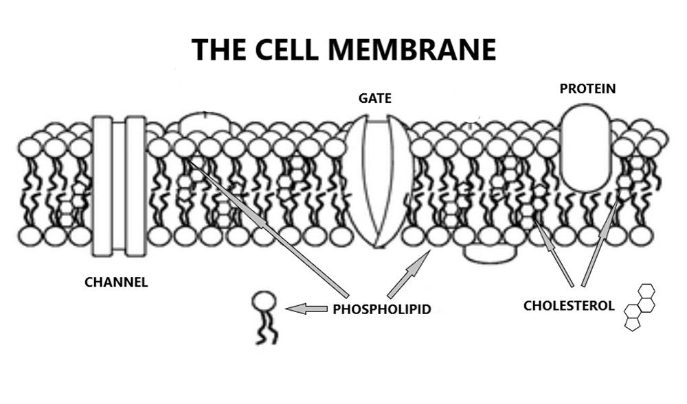
However, just to give you a couple of thoughts to consider. All blood vessels are lined by cells called endothelial cells. These cells, and indeed all cells in the body, have enormously complex mechanisms in place to regulate what gets into them, and what can then escape from them.

This regulation can control the movement of the largest molecule, down to the very smallest. Including tight control of the entrance, and exit, of single atoms e.g., sodium, or potassium, or calcium (in truth, in the body, these atoms exist as ions – which is just the name for a charged atom).

In short, nothing can gain entry to a cell unless the cell grants entry. Nothing [Unless you are a virus e.g., COVID19, whereby you hijack a receptor to sneak in – the cunning little devils]. For anything to gain entry, the cell must open a channel of some sort i.e., an ion channel. Or it has to manufacture a receptor. The receptor will then lock onto the substance floating in the bloodstream and drag it through the cell membrane and into the cell.

Just to pluck an example from the air. In order to get low-density lipoprotein into a cell, the cell must first synthesize a low-density lipoprotein (LDL) receptor. It then transports the receptor to the cell membrane. Once in place, it locks on to a passing LDL molecule, and then drags the entire LDL/receptor complex back inside – before breaking it down. Without this machinery getting to work, there is no way to get LDL into a cell. None.

Just to repeat this, from a slightly different direction. Cells can control the movement of single ions/atoms through their cell membranes. An LDL molecule is massive in comparison to an ion. Think rowing boat vs. super tanker.

Yet, if we are to accept the LDL hypothesis, we are supposed to believe that cell membranes simply step aside, or part like the red sea, to wave through a super-tanker sized molecule.

It all gets more unlikely. The cell must then allow the LDL molecule to propel itself from one cell membrane to the other. A very clever trick for an inanimate molecule, with no means of propulsion. Looked at from a human scale, if I were an LDL molecule, a cell would be about half a mile across.

Even if the LDL managed this, once it reached the other side of the cell, the LDL would need to pop back out of the cell and into the artery wall behind – using an inside-out LDL receptor perhaps?

Some people have therefore suggested the LDL molecules simply sneak between endothelial cells. But this, too, is impossible. Endothelial cells are tightly bound to each other, with a whole series of protein bridges a.k.a. ‘tight junctions.’ These too, can prevent the passage of single ions. Which means that nothing can get from the blood and into the artery wall though a side entrance either:

‘Tight junctions prevent the passage of molecules and ions through the space between plasma membranes of adjacent cells, so materials must actually enter the cells, in order to pass through the tissue.’1

If this were not the case, if the ‘endothelium’ did not represent a perfect barrier, we would all die immediately. To quote from a paper in the journal ‘Tissue Barriers’ yes indeed, there is such a journal:

Physiological barriers provide the framework for a boundary between circulating blood and interstitial fluid, a pre-requisite for mammalian life.’2

So, yes, there is simply no way for LDL to sneak between cells either.

Just to give you one example of what happens if you loosen the tight junctions between cells, we can look at the Ebola virus. This virus opens up the tight junctions between endothelial cells. At which point the endothelial barrier function is critically compromised, and blood can now escape into the tissues, and organs.

This leakage is the reason why Ebola is also known as a form of haemorrhagic fever. Blood appears in the cornea, so your eyes become bloodshot. You cough up the blood that that has escaped into your lungs. Your tongue falls off. Your urine is filled with blood from your kidneys, your bowel motions turn black, and then…. Bang, dead, from massive fluid loss and vascular collapse.

Ebola demonstrates very clearly the critical importance of tight junctions between cells. Open them up… and you die. How did you think Ebola actually kills you? I imagine that you, like me, before I started looking at this area in detail, probably never really thought much about it. You just vowed never to catch it:

Ebola patients experience a breakdown in endothelial barrier integrity that leads to massive fluid losses and vascular collapse.’3 

That would be where the ‘pre-requisite for mammalian life’ comes in.

Anyway, forgetting about that for the moment. Let me take you back to think about an alternative process model. To ask, so what is the disease process that allows you link smoking to, say, systemic lupus erythematosus? Because both things are most certainly causal. ‘A’ cause, rather than ‘the’ cause, of course.

And what of the many, many, other factors, not mentioned in Qrisk3? This is a very long list indeed. For example: cocaine use, antiphospholipid syndrome, Avastin, use of steroids, periodontitis, Sjogren’s syndrome, sickle cell disease, high fibrinogen levels. I could go on… for a few pages.

The harder you look, the more stones you unearth. In the end I had this massive pile of stones which became my two-thousand-piece jigsaw puzzle – just to change metaphors abruptly. A puzzle that didn’t even have a box to go with it, so I didn’t know what the picture was supposed to look like once I fitted all the pieces together. Was it a cow, or a lake, or a mountain scene?

At this point, you may get some insight as to why I called the book ‘the enduring mystery of heart disease’. Admit defeat …me? Well, usually, I am quite good at chucking in the towel. If at first you don’t succeed, give up, why make an idiot of yourself. But for some reason I did not, could not.

This bloody thing kept nagging away. I would lie awake at night considering the morphology (structure) of plaques. I contemplated the blood clotting system in all of its massively complex detail. I read papers outlining the thirty-two different types of HDL (good cholesterol). I discussed plaques with cardiologists and pathologists. I stumbled across entirely new entire worlds of research that I didn’t know existed. There were few rabbit holes down which I did not disappear.

The answer, when it finally lodged in my brain, was surprisingly simple. Not only was it simple, but it has also been kicking around for very nearly one hundred and seventy years. In 1852 Karl von Rokitansky published his ‘encrustation hypothesis.’ Describing what he saw when he closely examined atherosclerotic plaques.

‘Rokitansky proposed that the disease is the result of an excessive intimal deposition of blood components (blood clots) including fibrin. He maintained that localized thickening, atheromatous changes and calcification of the arterial wall are due to the repeated deposition of blood elements and their subsequent metamorphosis and degeneration on the lining membrane of the vascular wall.

There you go, that’s it. Blood clots, blood clots… and more blood clots.

In fact, I am being slightly disingenuous here. I had been thinking about blood clots for many years, but in a rather uninformed and directionless way. Many moons ago, when I was a medical student at Aberdeen, I was briefly taught cardiology by Dr Elspeth Smith. In a small group tutorial, she mentioned to us fresh faced students that LDL cannot penetrate the endothelium. Bing!

This was almost an off the cuff remark. Almost… but the way she said it… I knew that there was something critical here. A secret never to be told. Or, it was like a good murder mystery, where a chance remark represents the most important clue. My antennae pricked up at that moment and have not stopped twitching since.

She also wrote this over forty years ago:

‘After many years of neglect, the role of thrombosis (blood clots) in myocardial infarction is being reassessed. It is increasingly clear that all aspects of the haemostatic [blood clotting] system are involved: not only in the acute occlusive event, but also in all stages of atherosclerotic plaque development from the initiation of atherogenesis to the expansion and growth of large plaques.’

Yes, she knew what causes cardiovascular disease. I now know, for sure, that she knew. She never knew that I knew she knew. I was daft and did not take the opportunity to discuss things with her. Too much ego, I suppose. I also went off in other daft directions for many years. Looking for ‘the’ cause: the lost chord, the Ark of the Covenant, the Holy Grail. Of course, I never found ‘the’ cause, because it does not exist. Oh, well.

It was only when I was directed to return to process, that I came to fully recognise her genius – and the genius of others. I only hope that this book will help to raise her status up to where it belongs. Along with Rokitansky, Duguid, Ross and all those who pursued the blood clotting route over the decades, nay, centuries now.

Yes, I know this probably sounds very simple, and possibly completely wrong. There is no way you are going to believe straightaway that atherosclerotic plaques are just blood clots, ‘in various stages of metamorphosis and degeneration’. How can this possibly be true?

The reason why it can be true, is because it fits all the known facts about cardiovascular disease, and atherosclerotic plaques, and heart attacks, and strokes, and suchlike. Yes, everything.

For example. It is widely known, and accepted, that the single most common ‘final’ event in cardiovascular disease is the formation of a large blood clot – almost inevitably on top of an existing plaque. This can fully block an artery, leading to downstream mayhem.

It is also widely accepted that thrombus formation, on top of an already existing plaque, can make plaques suddenly jump in size. Here, for example, is a passage from a paper in the journal Atherosclerosis. This is about as mainstream a publication as you can get in cardiovascular disease research. The paper was called: ‘The role of plaque rupture and thrombosis in coronary artery disease.

In addition, plaque rupture and subsequent healing is recognized to be a major cause of further rapid plaque progression.’ 4

I could find a million quotes confirming this mechanism, without even breaking sweat.

In short, it is not remotely controversial to say that blood clots represent the final event, the thing that kills you. Nor is it controversial to say that plaques grow through the deposition of a new clot on top of an existing plaque. I would get very little argument from any cardiologist about this sequence of events. I know, I have had many such discussions.

But what is not accepted, will simply not be accepted, is that the arrival of a blood clot on the ‘healthy’ arterial wall is what initiates atherosclerotic plaque formation. Instead, it is almost universally accepted that it is LDL that does this, and LDL alone. Therefore, current conventional wisdom is that we have a three-step process:

  • The plaque starts – due to LDL leaking through the artery wall
  • The plaque grows – due to deposition of new blood clots on top of the existing plaque (which has become a point of clotting vulnerability)
  • A ‘final’ obstructive blood clot forms on the already grown plaque, causing a heart attack, or stroke – due to a blood clot

In one way, all I have done in this book is to make it clear that, yes, we do have a three-stage process. However, low-density lipoprotein plays no part in it. It is entirely driven by blood clots, from start to finish. Which means that the first step is not:

  • The plaque starts – due to LDL leaking through the artery wall

The first step is that:

  • The plaque starts, due to endothelial damage, and resultant clot formation (creating the ‘focus’ for further plaque growth)

Then, everything else follows, as described earlier by Elspeth Smith:

It is increasingly clear that all aspects of the haemostatic [blood clotting] system are involved: not only in the acute occlusive event, but also in all stages of atherosclerotic plaque development from the initiation of atherogenesis to the expansion and growth of large plaques.

Yes indeed, the Clot Thickens.

In truth, this is not a major leap in thinking? All I actually needed to do was remove low density lipoprotein from the picture. Once I did that, the jigsaw actually fitted together. Leave it in, and you cannot create a coherent hypothesis. Everything is distorted, nothing works, the picture is a mess. Take it out and it all makes sense.

I hope this brief foray into the process of cardiovascular disease has left you suitably enthused to buy the book, read it, and find out in much greater detail what is really going on here. And also, what you can do to prevent heart disease. It has been my life’s work. I hope I am right. I hope you can learn from it and enjoy it.

1: https://en.wikipedia.org/wiki/Tight_junction

2: https://www.ncbi.nlm.nih.gov/pmc/articles/PMC4836485/

3: https://www.sciencedirect.com/science/article/pii/S120197121500106X 4: https://www.sciencedirect.com/science/article/pii/S0021915099004797

The Clot Thickens is available in paperback and eBook.

Please support independent publishers and the author by purchasing a copy
Direct from the publisher
(UK SHIPPING ONLY)

Available online in paperback and eBook worldwide through Amazon:
UK | USA | DE | FR | ES | IT | JP | CA | AU

Additionally as eBook:
NL | BR | MX | IN


Let’s talk about C – just you and me

22nd October 2021

Studying cardiovascular disease for over thirty years can take you to some very interesting and seemingly strange places. Places where I never expected to find myself. Connections appear where you least thought they would be, and entirely new worlds of research open up. Very often, into places where mainstream medical thinking simply does not go.

The fascinating thing is how so many things end up looping back round, in ways that you would never have considered. One of those places is within the world of vitamins. Vital…amines, and the connections to cardiovascular disease.

Unfortunately, mainstream medicine has firmly locked vitamins into a tightly constrained box. Yes, it is accepted that vitamins are vital, because you die without them – that is where the ‘vital’, in vitamins, comes from after all.

However, it is universally believed that the exact requirements for all vitamins – known as the recommended daily allowance (RDA) – which were established decades ago. It is also universally believed that everyone gets sufficient vitamin intake from their diet, so there is absolutely no need for supplementation.

But how true are these comfortable assumptions? At one point I looked into Vitamin B12 deficiency. Even mainstream medicine agrees that this can, and does, occur and can lead to very serious medical problems. Irreversible nerve damage and paralysis, for example.

The normal range for Vitamin B12 in the UK varies from 190 to 950 picograms/ml (pg/ml).  [A picogram (pg) is one trillionth of a gram]. This at least is the normal range, in some laboratories… in some places the UK. In truth, it is almost impossible to find a consistent figure.

This ‘normal’ range also varies enormously from country to country. In Japan, for example, it is 500 – 1300 pg/ml. Thus, the lower range is nearly three times higher in Japan than the UK1. If you went to your GP in the UK with a level of four hundred and said you were Vitamin B12 deficient they would point you to the door. In Japan, they would treat you.

Another thing to note here is that the range is almost always ridiculously wide. It can vary by a factor of five! This alone suggests that people are pretty much guessing at what “is” normal. Despite this, most doctors remain perfectly content that a normal/healthy level has been well established and is based on robust science. There is no need to look again.

This is not the case, not even remotely. Here, for example, are the first three key recommendations on determining what constitutes Vitamin B12 ‘deficiency’ from the British Society for Haematology – with some of the highly technical text removed. They use the term cobalamin here, not Vitamin B12, although it is (basically) the same thing [Cobalamin (Vitamin B12) comes in several different formulations e.g., hydroxycobalamin, methylcobalamin, cyanocobalamin]

  • The clinical picture is the most important factor in assessing the significance of test results assessing cobalamin status because there is no ‘gold standard’ test to define deficiency
  • Serum cobalamin currently remains the first‐line test… Serum holotranscobalamin has the potential as a first‐line test, but an indeterminate ‘grey area’ may still exist.
  • Definitive cutoff points to define clinical and subclinical deficiency states are not possible, given the variety of methodologies used and technical issues, and local reference ranges should be established2

There you are, clear…. as mud. What the British Haematology Society informs us is that: there is no gold standard test for vitamin B12 deficiency, there are also ‘grey’ areas, and ‘definitive cut-off points to define deficiency states are not possible.’

As with almost all areas of medical research, the more you dig down, the more uncertain things become. The authors of the report are not even sure if you should be measuring cobalamin, or holotranscobalamin – whatever that may be. It sounds like something from Star Trek.

When it comes to vitamin D there is a similar lack of clarity. In the UK, recent guidelines on vitamin D suggested more people should take supplements… finally. However, the NHS advice on vitamin D then goes on to make this statement:

‘…although roughly one in five people has low vitamin D levels, this is not the same as a vitamin D deficiency. It is not accurate to say that millions of people are at risk of deficiency.’3

So, according to the NHS, a low level is not a deficiency. However, a low level cannot, by definition, be normal. For, if it were normal, you could not call it “low”! So, what is it? Here is where words really start tripping over each other. Low, deficient, normal…optimal, inadequate, sub-optimal? Trying to pin any clear definition down is, I can assure you, like trying to pick up mercury using your fingertips.

In the same article it is stated that a blood level of 20nmol/l is ‘sufficient’. Again, what does sufficient mean? Is it the same thing as normal, or is it optimal? Is this really the level we should be aiming for? The National Institutes of Health in the US provides completely different figures for vitamin D deficiency. Or, as they choose to call it – ‘inadequacy.’

‘Some people are potentially at risk of inadequacy at 30 to 50 nmol/L (12–20 ng/mL). Levels of 50 nmol/L (20 ng/mL) or more are sufficient for most people. In contrast, the Endocrine Society stated that, for clinical practice, a serum 25(OH)D concentration of more than 75 nmol/L (30 ng/mL) is necessary to maximize the effect of vitamin D on calcium, bone, and muscle metabolism.

The truth is that, wherever you look the figures are all over the place. Made more complicated by the fact that the US uses different units of measurement to everyone in the civilised world, by which I mean Europe… of course. When they say seventy-five, they really mean thirty. ‘You say nanograms per millimole, I say nanomoles per litre – let’s call the whole thing off.

So, should you be aiming for 20nmol/l? Or is it thirty or forty, or fifty? If in doubt aiming higher would be my advice, better safe than sorry. After all, it has been found that the majority of people with cancer have ‘low’, if not ‘deficient’ levels of Vitamin D.

‘More than three-fourths of people with a variety of cancers have low levels of vitamin D, and the lowest levels are associated with more advanced cancers, a new study suggests.’4

If having a low level of Vitamin D means you are more likely to get cancer, then I would certainly define this as deficient, not low, and I would certainly want to do something about it.

And it is not just cancer. There are studies linking low vitamin D to many other diseases, such as kidney disease and, more importantly for the sake of this article, diabetes, and cardiovascular disease. As highlighted in this paper: ‘Vitamin D deficiency increases risk of nephropathy and cardiovascular diseases in Type 2 diabetes mellitus patients.’

‘Vitamin D (VD) deficiency is associated with insulin function and secretion. It is linked with diabetes mellitus (DM) progression, and complications were also recorded…The evidence from this study suggest that patients with Type 2 diabetes with vitamin D deficiency are at higher risk for developing CVD and nephropathy [kidney damage].5

Additionally, a low Vitamin D level can be a factor that drives obesity and diabetes in the first place. Here, from the study: ‘Vitamin D deficiency is a risk factor for obesity and diabetes type 2 in women at late reproductive age.’

‘Our results showed that vitamin D insufficiency is highly prevalent in the population of healthy women. Low 25(OH)D [a form of vitamin D] levels correlated with high body fat, glucose levels and decreased insulin sensitivity. We conclude that vitamin D deficiency is a potential risk factor for obesity and development of insulin resistance leading to diabetes type 2. 6  

If you choose to look, the evidence for the potential harms of low… deficient… inadequate…insufficient… suboptimal Vitamin D stretch on, and on, and on. Cancer, diabetes, obesity, kidney failure, cardiovascular disease. In the era of COVID19, there are also significant benefits from vitamin D in boosting of the immune system and reducing the risk of infection.7

Cutting to the chase, most doctors are eager to dismiss the benefits of Vitamin D on, pretty much, anything. However, I think the evidence for benefits on overall, and cardiovascular health are overwhelming.

Of equal importance is the fact that vitamin D is incredibly safe to take. It is true that toxicity has been seen in a few people, very few. However, it took sixty thousand units a day for several months to reach this point.

‘Taking 60,000 international units (IU) a day of vitamin D for several months has been shown to cause toxicity.8

Frankly, taking that dose would be nuts, and would not be required by anyone, ever. Personally, I take nine thousand units a day from October to March. I am l looking to get my levels above 50nmol/l, and keeping them there, if possible.

And why on earth would you not?  Vitamin D is remarkably safe, and cheap. In the summer you don’t need to take any at all. You can get all you need by going out in the sun when it shines and, shock, horror, exposing your skin for an hour or so. Even more if you like.

Why do doctors dislike vitamins so much? It is complicated, but primarily driven by the pharmaceutical industry, who absolutely hate the idea of people buying ‘health’ products that they cannot make any money from. So, they make wild claims about vitamins damaging health, and suchlike. They are simply trying to take out the opposition, usual tactics. For example:

‘….a USA TODAY investigation finds that a wide array of dietary supplement companies caught with drug-spiked products are run by people with criminal backgrounds and regulatory run-ins. Consumers buying products from these firms are in some cases entrusting their health and safety to people with rap sheets for crimes involving barbiturates, crack cocaine, Ecstasy and other narcotics, as well as arrests for selling or possessing steroids and human growth hormone. Other supplement company executives have records of fraud, theft, assault, weapons offenses, money laundering or other offenses, the investigation shows.9

Blah de blah. I amuse myself by reading the title of the book by Peter Gøtzsche: ‘Deadly Medicines and Organised Crime. How big pharma has corrupted healthcare.’ As he says. ‘If you don’t think the system is out of control, please email me and explain why drugs are the third leading cause of death… If such a hugely lethal epidemic had been caused by a new bacterium or a virus, or even one-hundredth of it, we would have done everything we could to get it under control…

Something reinforced by Richard Smith (previous long-time editor of the British Medical Journal) in the foreword to Gøtzsche’s book:

‘It is scary how many similarities there are between this industry (the pharmaceutical industry) and the mob. The mob makes obscene amounts of money, as does this industry. The side effects of organised crime are killings and death, and the side effects are the same in this industry. The mob bribes politicians and others, and so does the drug industry.’

Just in case you think Gøtzsche and Smith are over-reacting, here is what Harvard University states:

‘Few know that systematic reviews of hospital charts found that even properly prescribed drugs (aside from misprescribing, overdosing, or self-prescribing) cause about 1.9 million hospitalizations a year. Another 840,000 hospitalized patients are given drugs that cause serious adverse reactions for a total of 2.74 million serious adverse drug reactions. About 128,000 people die from drugs prescribed to them. This makes prescription drugs a major health risk, ranking 4th with stroke as a leading cause of death. The European Commission estimates that adverse reactions from prescription drugs cause 200,000 deaths; so together, about 328,000 patients in the U.S. and Europe die from prescription drugs each year. The FDA does not acknowledge these facts and instead gathers a small fraction of the cases.10

Ouch. And there are those who think I am highly critical of the pharmaceutical industry. I’m a pussy cat in comparison. How many deaths have there been from vitamins? Last time I looked; it was one, over a ten-year period. I think a large crate of vitamin D fell off a lorry and squashed someone… (joke).

Anyway, yes, as you may have noticed, I have not yet talked about C… Vitamin C. I have just been setting the scene. Rearranging the mental furniture. So, now to vitamin C. How much do you need? What good does it do?

I find it somewhat strange that almost all animals can synthesize their own Vitamin C, but we cannot. Along with a few great apes, a couple of fruit bats and guinea pigs. Animals synthesize it from glucose, in four steps.

Humans have retained the first three steps but lack the fourth. We lost this fourth step about forty million years ago. Perhaps because we learned to re-cycle vitamin C within our red blood cells, so we need far less of it. The ‘electron transfer hypothesis.’ If making Vitamin C uses up resources that we need for other things… why bother. Just eat it, there is plenty about11.

Anyway, for whatever the exact reason, we lost the ability to make vitamin C. So, we now have to eat it. Mostly from fruit and plants. Tricky if you are Inuit. However, animal meat does contain enough vitamin C to keep the Inuit going.

There is a hypothesis that the Inuit ensure that they eat the adrenal glands of various animals they kill, because this is where there is the highest concentration of vitamin C lies. I don’t think I have seen this proven. Anyway, how could the Inuit possibly have known where vitamin C was concentrated? A clever trick indeed. Do they have secret biochemical labs hidden within glaciers?

Moving on. What happens if you do not eat enough vitamin C? Well, a whole lot of different things. But the most serious problem is that vitamin C is required to create collagen. Think of collagen as being like the steel bars in concrete, providing support and strength for tissues around the body. Without collagen, things can start to break apart quite dramatically.

Blood vessels, for example, need a lot of collagen, as they have to withstand a lot of pressure, and squeezing and bending and suchlike. So, one of the first clinical signs of scurvy (Vitamin C deficiency) is often bleeding gums. Followed by bleeding everything else. Followed by bleeding to death. Not recommended.

What is both pertinent, and fascinating at this point in the vitamin C story, is that evolution came up with a plug to reduce the risk of bleeding to death in vitamin C deficiency. Until enough vitamin C could be found and consumed again, and collagen synthesis got back to normal.

This plug is called Lipoprotein(a). Or Lp(a).

If blood vessels start to crack, this action attracts a Lp(a) to the scene. It then flings itself at the cracks, to form a plug that is highly resistant to being broken apart. More so, than any other part of a blood clot. It achieves this resistance by using a very clever trick, which is that it blocks the activation of the enzyme specifically designed to break down blood clots.

At this point I need to explain a bit more about blood clots… So, off we go once more, on a detour.

The enzyme designed to break clots apart is plasmin, which does the job of slicing apart strands of fibrin. Fibrin is the very tough strand of protein that wraps around all blood clots, then binds them together, then tightens up the entire clot up and makes it very tough and difficult to ‘lyse’ i.e. slice apart.

Fibrin is constructed when smaller pieces of protein, called fibrinogen are linked up, end to end, to form the much longer fibrin strand. This is the final step of the monstrously complex ‘clotting cascade’. [You could not allow long strands of fibrin to float about freely in the blood. They would just end getting tangled around everything else and getting stuck in various vital places.]

So, whilst fibrin has a critical function in blood clot formation, if you cannot break it down – once the bleeding has stopped, and repair has started – then you cannot break apart the blood clot either – at least not easily. And if you cannot break apart blood clots, then they are going to hang around – almost forever. Which is not a good thing, as you can probably imagine.

Which is where the enzyme known as plasmin comes in. Once bleeding has stopped, plasmin is ‘activated’ to slice – or lyse – the clot apart, and then it is gone.

How do you activate plasmin? Well, this process starts with another protein called plasminogen – which is incorporated into all blood clots as they form. Plasminogen then sits there doing nothing much. However, you can convert plasminogen into plasmin using another enzyme called tissue plasminogen activator (TPa).

TPa + plasminogen → plasmin → fibrin sliced apart ‘lysed’

Yes, step after step… after step. Tissue plasminogen activator is now made commercially and is colloquially known as a ‘clotbuster’. It is often given to people having a stroke to ‘bust’ the clot apart. [Unless you are having a stroke due to a bleed, not a clot, at which point given TPa would not be a great idea].

So, and keep holding on here, because I am going to get back to Vitamin C in a bit… so, what if you could not break up clots? At least, not so easily. Well, whilst this is a good thing if your blood vessels are cracking due to a lack of collagen, as the blood clot ‘plugs’ will need to last for a long time. At least until Vitamin C intake goes up, and collagen can be made.

However, if you do not have scurvy, having blood clots that resist lysis is a bad thing, because these clots are more likely going to hang around for ages. They will be stuck to blood vessel walls for quite a long time. Which means that they can become the focus for atherosclerotic plaques. [At least this is what happen if you believe in the thrombogenic hypothesis – which I do].

Now, getting back to Lp(a). How does it stop clots being broken down? Well, the(a) in Lp(a) stands for apolipoprotein(a). This protein is almost identical to plasminogen – the protein that is incorporated into all blood clots as they form. However, apolipoprotein(a) cannot be converted to plasmin by tissue plasminogen activator. Instead, it acts as a tissue plasminogen activator inhibitor. It jams up the active site of TPa.

So, deep breath. If you have a lot of Lp(a) around, you are in danger of creating difficult to shift blood clots. As outlined in the paper ‘Lipoprotein(a) as a modifier of fibrin clot permeability and susceptibility to lysis.’

‘We here provide the first evidence that elevated plasma Lp(a) levels correlate with decreased fibrin clot permeation and impaired susceptibility to fibrinolysis both in apparently healthy subjects and patients with advanced coronary artery disease. The relationship between Lp(a) and clots …are associated with extremely unfavourable clot properties12.

Therefore, if you have a lot of Lp(a) in you blood, you will have blood clots with ‘extremely unfavourable clot properties.’ And so, you may end up dying of cardiovascular disease. Here is an article from the New York Times:

‘To millions of Americans, Bob Harper was the picture of health, a celebrity fitness trainer who whipped people into shape each week on the hit TV show “The Biggest Loser.”

But last February, Mr. Harper, 52, suffered a massive heart attack at a New York City gym and went into cardiac arrest. He was saved by a bystander who administered CPR and a team of paramedics who rushed him to a hospital, where he spent two days in a coma.

When he awoke, Mr. Harper was baffled, as were his doctors. His annual medical checkups had indicated he was in excellent health. How could this have happened to someone seemingly so healthy?

The culprit, it turned out, was a fatty particle in the blood called lipoprotein(a). While doctors routinely test for other lipoproteins like HDL and LDL cholesterol, few test for lipoprotein(a), also known as lp(a), high levels of which triple the risk of having a heart attack or stroke at an early age.

You may think, why have I never heard of Lp(a). Fear not, you are not alone, as most doctors have never heard of it either. The surprising fact is that, although you may think you have never heard of Lp(a) you have. Because it is actually….

Drum roll, great suspense…

It is…. Low Density Lipoprotein (LDL). Yes, it is ‘bad cholesterol’ itself. The evil substance of doom itself. What a remarkable coincidence…

You think I am pulling your leg. I am, but only slightly. In fact, to be fully accurate, Lp(a) is actually low-density lipoprotein (LDL), with an extra strand of protein attached to it. And that protein is apolipoprotein(a).

Yes, apolipoprotein(a), the very protein that pretends to be plasminogen. The protein that inhibits blood clots from being broken apart. This is all a bit like a Sherlock Holmes story. Ladies and Gentlemen, I give you…

‘The tragic case of mistaken identity.’

‘I put it to you sir, that when you looked at atherosclerotic plaques and saw LDL within them, you were actually looking at Lp(a) molecules, but you did not recognise them. Because you miserably failed to look for the apolipoprotein(a).’

Or, to switch metaphors in a heavy-handed manner to Cluedo.

‘It was Lp(a) wot done it, in the left anterior descending artery, with an apolipoprotein(a) molecule.’

Or, to put it more technical speak, from the paper ‘Quantification of apo[a] and apoB in human atherosclerotic lesions.’:

These results suggest that Lp[a] accumulates preferentially to LDL in plaques, and that plaque apo[a] is directly associated with plasma apo[a] levels and is in a form that is less easily removable than most of the apo B. This preferential accumulation of apo[a] as a tightly bound fraction in lesions, could be responsible for the independent association of Lp[a] with cardiovascular disease in humans13.’  

Oh, my goodness, it is all so very complicated, is it not? Well, it is both complicated, and fascinating. You start looking at Vitamin C, and you end up comparing the molecular structure of plasminogen and apolipoprotein(a). Then you find that Lp(a) is, to all intents and purposes, LDL.

Now, let me see. Where does vitamin C properly fit into this tale?

Well, if you don’t have enough vitamin C, then you are more likely to end up with cracks in your blood vessels. These cracks will then be plugged by small blood clots, containing a lot of Lp(a). If you have a high Lp(a) level, then these small blood clots will be even bigger, and even more difficult to remove.

Which means that if you have a high Lp(a) level, it would be a splendid idea to ensure that you never become vitamin C deficient. Indeed, even if you do not have a high Lp(a) level it would be a splendid idea to ensure that you do not become vitamin C deficient. Because cracks in blood vessel walls are never a good thing. Ending up, potentially, as the focus for atherosclerotic plaques.

Linus Pauling, a famous double Nobel Prize winner, believed that ‘sub-clinical’ Vitamin C deficiency was ‘the’ cause of cardiovascular disease. He also believed that if everyone took enough vitamin C, cardiovascular disease would disappear.

Personally, I do not think it is ‘the’ cause of cardiovascular disease, but I do think that it is ‘a’ cause. That is, whether or not you have a high Lp(a) level. Of course, a high Lp(a) level is likely to make things far worse, were you to end up vitamin C deficient.

However, the vitamin C, cardiovascular disease story does not end here. Because vitamin C has many other critical functions that link back to cardiovascular disease in one way, or another. For example, it has a more general function in protecting endothelial cells from harm, and supports the integrity of the vascular system. Here, from the paper ‘Role of Vitamin C in the function of the vascular endothelium’:

‘Vitamin C, or ascorbic acid, has long been known to participate in several important functions in the vascular bed in support of endothelial cells. These functions include increasing the synthesis and deposition of type IV collagen in the basement membrane, stimulating endothelial proliferation, inhibiting apoptosis (endothelial cell death), scavenging radical species, and sparing endothelial cell-derived nitric oxide to help modulate blood flow. Although ascorbate may not be able to reverse inflammatory vascular diseases such as atherosclerosis, it may well play a role in preventing the endothelial dysfunction that is the earliest sign of many such diseases.’

Supplementation to upper normal plasma ascorbate levels is clearly indicated in most diseases and conditions in which ascorbate is depleted. However, it is seldom a priority, because patients, physicians, and health authorities are unaware of the increasing evidence for multiple potentially important functions of ascorbate. With regard to the endothelium, it is worth emphasizing observations made more than 50 years ago that early scurvy generates endothelial disruption in guinea pigs, which resembles atherosclerosis and is fully and rapidly reversible with ascorbate repletion.14

Yes, with regard to the last part about guinea pigs. Many years ago, a researcher deliberately made guinea pigs ‘scorbutic’ – the medical term for the state of vitamin C deficiency, a.k.a. scurvy. At which point they developed atherosclerotic plaques.

When the vitamin C was added back into their diet, the atherosclerotic plaque disappeared. [Unless you left it too long, in which case, the plaques remained]. Best animal experiment on atherosclerosis ever done – never repeated.

Having just said all of this. I do not believe that most of us, most of the time, are lacking vitamin C – to any degree. At least I do not think so. However, if we become infected – with almost anything – the requirement for vitamin C shoots up. Because Vitamin C gets burned up protecting the endothelium, and it also supports the immune system

‘The role of vitamin C in lymphocytes is less clear, but it has been shown to enhance differentiation and proliferation of B- and T-cells, likely due to its gene regulating effects. Vitamin C deficiency results in impaired immunity and higher susceptibility to infections. In turn, infections significantly impact on vitamin C levels due to enhanced inflammation and metabolic requirements.15

Another key thing to know about vitamin C, again closely related, is that people with type II diabetes, and people who smoke, have reduced circulating levels of Vitamin C.

‘Although T2DM [type II diabetes mellitus] is not traditionally considered a risk factor for vitamin C deficiency, our research indicates that those with prediabetes or T2DM are more likely to have inadequate or deficient plasma vitamin C concentrations. This did not appear to be due to a lower dietary vitamin C intake, so dietary advice needs to emphasise the importance of consuming high vitamin C foods.16

What links smoking, type II diabetes, and vitamin C? Here I am hypothesizing a little. What links them is that with smoking, and type II diabetes, the endothelium is under ‘attack’. High blood sugar levels damage the glycocalyx (the protective lining of endothelium), and so do the nanoparticles that enter the bloodstream if you smoke.

Here is a quote from the paper: ‘Loss of endothelial glycocalyx during acute hyperglycemia coincides with endothelial dysfunction and coagulation activation in vivo.’ (In vivo means in a real live person, not just in vitro – in a test tube). Jargon alert:

‘Hyperglycemia is associated with increased susceptibility to atherothrombotic stimuli. The glycocalyx, a layer of proteoglycans covering the endothelium, is involved in the protective capacity of the vessel wall. We therefore evaluated whether hyperglycemia affects the glycocalyx, thereby increasing vascular vulnerability…

In the present study, we showed that the glycocalyx constitutes a large intravascular compartment in healthy volunteers that can be estimated in a reproducible fashion in vivo. More importantly, we showed that hyperglycemic clamping elicits a profound reduction in glycocalyx volume that coincides with increased circulating plasma levels of glycocalyx constituents like hyaluronan, an observation that is consistent with the release of glycocalyx constituents into the circulation17.’

Looking specifically at smoking:

‘Vascular dysfunction induced by smoking is initiated by reduced nitric oxide (NO) bioavailability and further by the increased expression of adhesion molecules and subsequent endothelial dysfunction. Smoking-induced increased adherence of platelets and macrophages provokes the development of a procoagulant and inflammatory environment.18 

Essentially, smoking and high blood glucose both damage the endothelium, which results in low vitamin C levels, as the endothelial cells burn through Vitamin C to maintain themselves. Ergo, fi you have type II diabetes, or smoke, you need more Vitamin C to maintain healthy levels….

Now, before I introduce you to far too many new and difference concepts – I did mention everything starts linking back together in completely unexpected ways – it is time to draw our little tale of Vitamin C together.

The first thing to say about vitamin C is that it is vital. I have only covered a few of the essential functions that it has in the human body. Those most closely related to cardiovascular disease. Importantly, it is almost impossible to cause harm by overconsumption. It is just about as safe to take, as anything can possibly be.

The next thing to say is that most of us, most of the time, probably have sufficient vitamin C intake, and require no supplements.

However, if you have a high Lp(a) level, then any damage caused by a lack of vitamin C will be amplified, dure the fact that Lp(a) sticks very tightly to areas of endothelial damage, making the resultant blood clot very difficult to remove. So, for those with high Lp(a) levels, I would recommend one gram of vitamin C a day – forever.

If you smoke, or have diabetes, the lining of your artery walls (glycocalyx and endothelium) are under constant attack from nasty substances – smoke nanoparticles and high blood glucose. This, too, will create ‘cracks’ in blood vessels.

In both situations Vitamin C is also used up more rapidly, trying to protect against this damage. So your vitamin C level is likely to be low. Which means that you too, should take one gram of vitamin C a day – forever. [Or you could try stopping smoking]

In addition, in many infections, the endothelium is under severe attack. Either directly from the microorganisms entering and killing endothelial cells (see under COVID19), or from the exotoxins (toxic waste products) released by any bacteria in the bloodstream.

The most severe endothelial attack occurs in sepsis (infection of the blood), where the exotoxins strip away the endothelium, resulting in widespread blood clotting (disseminated intravascular coagulation DIC). Which is the thing that, primarily, kills you with sepsis.

Whilst sepsis represents an extreme situation, it is still the case that if you are suffering from an infection, of any sort (gingivitis or periodontal disease) your requirement for vitamin C will shoot up. Which means that you should take as much Vitamin C as you can tolerate. Up to ten grams a day. I cannot take this amount due to the impact it has on my gastrointestinal tract. Loose, is the word. Very loose. Looser than loose.

But if you can tolerate it, Vitamin C will help to protect your endothelium. It will also boost the functioning of your immune system.

So, there we are then, vitamin C. My second favourite vitamin, after vitamin D. In the winter I take a gram a day. Along with my nine thousand units of vitamin D. Almost all medics will instantly dismiss this as ‘woo woo’ nonsense. I would tend to argue that this is because they know absolutely nothing about vitamins, or their critical roles in human physiology, and have never bothered to find out.

Pop quiz for your doctor, next time you see them. Ask them how a lack of vitamin C causes scurvy. What is the primary disease process? Watch them scrabble to bring up a Google search. Then ask them about Lp(a). What it is, what it does… I guarantee that silence will be the stern reply.

1: https://advances.augusta.edu/1014#:~:text=Interestingly%2C%20in%20Japan%20the%20reference,methylmalonate%20levels%20are%20not%20checked.

2: https://onlinelibrary.wiley.com/doi/full/10.1111/bjh.12959

3: https://www.nhs.uk/news/food-and-diet/the-new-guidelines-on-vitamin-d-what-you-need-to-know/

4: https://www.webmd.com/cancer/news/20111004/low-vitamin-d-levels-linked-to-advanced-cancers#1

5: https://www.ncbi.nlm.nih.gov/pmc/articles/PMC6540771/

6: https://pubmed.ncbi.nlm.nih.gov/23924693/

7: https://vitamindforall.org/letter.html

8: https://www.mayoclinic.org/healthy-lifestyle/nutrition-and-healthy-eating/expert-answers/vitamin-d-toxicity/faq-20058108#:~:text=Advertisement&text=The%20main%20consequence%20of%20vitamin,the%20formation%20of%20calcium%20stones.

9: https://eu.usatoday.com/story/news/nation/2013/12/19/dietary-supplements-executives-criminal-records-spiked/4114451/

10: https://ethics.harvard.edu/blog/new-prescription-drugs-major-health-risk-few-offsetting-advantages

11: https://academic.oup.com/emph/article/2019/1/221/5556105

12: https://onlinelibrary.wiley.com/doi/full/10.1111/j.1538-7836.2006.01903.x

13: http://www.jlr.org/content/32/2/317.full.pdf

14: https://www.ncbi.nlm.nih.gov/pmc/articles/PMC3869438/

15: https://www.ncbi.nlm.nih.gov/pmc/articles/PMC5707683/

16: https://www.ncbi.nlm.nih.gov/pmc/articles/PMC5622757/

17: https://diabetes.diabetesjournals.org/content/55/2/480

18: https://www.ncbi.nlm.nih.gov/pubmed/24554606

Inclisiran sneaks through under cover of COVID19

23rd September 2021

With all medical eyes on COVID19, a cardiovascular drug with no proven benefit – at all – has been approved by NICE (The UK National Institute for Health and Care Excellence). Once a drug is approved by NICE it can, and will, be prescribed by doctors in England and Wales and Northern Ireland. Scotland has its own system.

NICE is also hugely influential beyond this small island. A NICE approval usually means a green light for approval in many other countries as well. Countries who assume that NICE will have carried out an in-depth ‘expert’ analysis using a set-up that they don’t have yet.

Which means that drug companies are always very keen to get NICE approval. It is a de-facto quality stamp. ‘This drug is both safe and cost-effective. You may now prescribe it everywhere in the world…’

Cost-effectiveness means that a drug does not just provide some clinical benefit. It must provide benefits that give you decent bang for your bucks. The ‘bang for your bucks’ measure used is the Quality Adjusted Life Year (QALY).

A QALY = one year of perfect health.

Of course, no healthcare intervention will ever give you one extra year of perfect health. Nothing is ever as clear cut as that. However, if you are suffering from a painful arthritic hip – and your quality of life is 50% perfect, or 0.5 – and you get a hip replacement, then your quality of life may rise from 0.5 to 0.9. So, you get 0.4 of a QALY/per year improvement.

After five years you have gained 0.4 (QALYs) x 5 (years) = 2 QALYs.

If the hip replacement operation has cost £10,000. The cost per QALY = £5,000. The cost per QALY obviously goes down if you live longer. That is a very simple example, most calculations become exceedingly complex. Measuring quality of life, for example, is fraught with difficulties.

In general NICE will approve a healthcare intervention if the cost per QALY is less than £30,000. This figure can never be pinned down. I often liken it to a blob of mercury. If you try to pick it up, it just slips, and slides, and fragments.

Indeed, this £30,000 figure never had any economic basis, or any other basis. It was simply plucked from the air because … well, because it seemed reasonable.

Here, from a discussion in the UK Parliament when NICE was first starting up:

‘There is clearly confusion about the cost per QALY threshold. Witnesses questioned whether there was any evidence to support the level that appears to be used. Professor Devlin told us that, “the threshold has no explicit basis or location in evidence”. Others agreed that it was “arbitrary”. Professor Smith confirmed…

Professor Rawlins admitted that the threshold was not based on “empirical research” as no such research existed anywhere in the world. He told us instead that the threshold was: …really based on the collective judgment of the health economists we have approached across the country. There is no known piece of work which tells you what the threshold should be.

No public discussion has ever taken place of the suitability of the threshold used. The American Pharmaceutical Group pointed out that the threshold has “never been the subject of public debate or Parliamentary approval. Cancer Research UK also argued that the threshold should be discussed openly and the reasons for its level should be determined in consultation with interested organisations.’ 1

I love it when people say things like ‘the threshold has no explicit basis or location in evidence.’ The short word – no – would have done nicely. As in, there is no evidence. Instead, we get the concept of no explicit basis or location in evidence. Listen guys, just get rid of the words: explicit, basis, or, and location. Why use five words when one will do?

Anyway, it has always amused me that NICE spends vast amounts of time and effort trying to establish with great accuracy whether a healthcare intervention meets a cost per QALY threshold… that was simply made up.

You might as well have got a cane with a hook on the end to pluck one of a thousand plastic yellow ducks floating in a pond with a random number written on the bottom. ‘Oooh look, it says thirty thousand… so that is the figure we shall use.’ Yes, really.

Anyway, as custom is king, this £30K figure – which has remained unaltered for twelve years [has anyone at NICE ever heard of inflation – or maybe a made-up figure cannot be affected by inflation] is unquestioned, and unquestionable. It is carved in stone. ‘And God did sayeth unto the multitude that thirty thousand pounds per QALY shalt be my law unto the end of time. Amen.’ Anyway, following that little history lesson, let us gaze upon the cost per QALY calculations that NICE used for Inclisiran – a new LDL lowering injection to be given twice a year. And below are the calculations [or at least the calculations that I could be bothered to copy]:

As you can see Inclisiran meets the cost per QALY criteria with ease. Well, actually, in truth, you cannot see anything because all the figures have been redacted. It amuses me further that NICE have decided that a table which contains no information of any use is ‘confidential’. Well, of course, it is not very confidential, because I can see it, and so can you, if you decide go to: https://www.nice.org.uk/guidance/gid-ta10703/documents/1

So, which part of that completely pointless table is confidential? It is clearly not confidential that it is confidential. Maybe this is some strange double-bluff. Perhaps you can scrape away the black areas to reveal the numbers beneath, and win a million-pound prize?

I suspect that written beneath them will be the phrases. ‘How much?’ ‘You’re having a laugh.’ Or ‘We will not release your family unless you pay this as ransom.’ And suchlike.

Enough of this. NICE is a public funded organisation that is supposed to work on our behalf. They have decided that Inclisiran is cost-effective, yet they will not let anyone see the figures that they used? You would think they would be shouting it from the rooftops. ‘Look how fantastic it is. Look at the size of that discount. Gaze with wonder on the magnificent cost-effectiveness of Inclisiran.’ No, instead, they are very shy about it. Like little meercats sensing danger and scurrying down into the darkness.

Without any figures, the NICE appraisal is essentially hundreds of pages of utterly meaningless guff. As I used to say, once upon a time, when teaching my children to count. ‘One two, miss a few, ninety-nine, one-hundred’.

How much are we paying for Inclisiran? Nobody knows. Because NICE won’t tell us. We know it should be/could be around £4,000/year ($5,000). So, some sort of discount has been negotiated. How much … well, that’s a secret. A secret. Why? In case we all rush round to Novartis headquarters and try and buy some at a bigger discount?

Secret or not, the truth is that I do not need to know the exact cost of Inclisiran. Because I already know that whatever it costs, the cost per QALY current stands at infinity i.e., ∞. I know this because, at present, there is no evidence that it provides any benefit, on any clinical outcome. By which I mean no evidence that it prevents strokes, heart attacks or, in fact, anything.

Based on this knowledge the Cost per QALY equation goes something like this:

Cost per year of Inclisiran/benefit in QALYs = cost per QALY

With Inclisiran let us set the cost per year at £4,000, and benefit at zero:

£4,000/ 0 = ∞ (infinity)

Let us set the cost at £1 and run the equation again

£1/0 = ∞ (infinity)

You see how simple it is to work out the cost per QALY when there is no benefit. It always ends up at infinity. If it cost one Turkish lira, the cost per QALY would still be infinity. I certainly did not need several hundred pages of guff to tell me this. You ought to try reading an endless NICE report sometime. My advice is, don’t bother.

Anyway, if you do read what NICE has to say about Inclisiran you will find the following key sentences in the NICE appraisal documentation.

‘There is also no long-term evidence on whether Inclisiran reduces cardiovascular events. This means the clinical evidence and the cost-effectiveness estimates are very uncertain.’2

Let me rephrase the first sentence.

THERE IS NO EVIDENCE THAT INCLISIRAN WORKS

Let me rephrase the second sentence

THIS MEANS, THE CURRENT COST-EFFECTIVNESS ESTIMATES ARE NONSENSE

Yes, it lowers LDL (low density lipoprotein), we do know that. We do not know if this will have an impact on cardiovascular deaths, or overall mortality. NICE assumes that if you lower LDL, you will reduce cardiovascular death – and suchlike.

However, this is not necessarily true. Repatha (evolucamab) is a drug which has exactly the same mechanism of action as Inclisiran but needs to be given every two weeks instead of twice a year. Both Inclisiran and evolucamab are PCSK9-inhibitors. [They block the breakdown of LDL-receptors in the cell, which means that more LDL receptors are available to pluck LDL molecules from the blood, thus reducing the blood levels]. Both drugs reduce the LDL level by pretty much the same amount.

In the FOURIER study on Repatha the results were the following

Cardiovascular mortality – total numbers:

Repatha          = 251

Placebo          = 240

Overall mortality

Repatha         = 444

Placebo         = 4263

Yes, Repatha lowered LDL to the same degree as Inclisiran and yet slightly more people died taking Repatha than died taking placebo. Repatha has been approved and launched, although you may wonder how or why.

In short, just because a drug lowers LDL does not mean it does any good. Just to give another example, the drug evacetrapib lowered LDL by 37% (and increased HDL by 132%). It, too, had absolutely no impact on cardiovascular mortality.

‘Although the cholesteryl ester transfer protein inhibitor evacetrapib had favorable effects on established lipid biomarkers, treatment with evacetrapib did not result in a lower rate of cardiovascular events than placebo among patients with high-risk vascular disease.’ 4 

Just in case you are wondering, Evacetrapib did not launch. Nor did another three drugs in the same class that all had ‘favourable effects on established lipid biomarkers’ but achieved nothing. One of them, torcetrapib, increased cardiovascular death by 50%.

In short, approving drugs, or launching drugs before you have any evidence that they do anything – other than having a favourable effect on an established lipid biomarker – is ridiculous. But never mind, longer term studies on Inclisiran will be completed by 2023, and 2026. When will they actually be published?

Who cares, by the time they are published, Inclisiran will have made billions, and no-one will care if the results are positive, or negative, as it will have become established as ‘standard’ treatment.

A number of us found the NICE approval of Inclisiran so ridiculous that we wrote them a letter. (See below). I do not imagine it will have the slightest impact.


To: Sharmila Nebhrajani OBE,

Chair: National Institute for Health and Care Excellence

2nd Floor, 2 Redman Place

London E20 1JQ

cc. The Right Honourable Sajid Javid, MP Secretary of State for Health and Social Care Department of Health

Richmond House 79 Whitehall London, SW1A 2NS

15th Sept 2021

Concerns about the latest NICE draft guidance on Inclisiran

Introduction:

We are concerned about your draft final guidance recommending the novel anti-cholesterol drug inclisiran (Leqvio and made by Novartis) for people with primary hypercholesterolaemia or mixed dyslipidaemia who have already had a cardiovascular event such as a heart attack or stroke.

We would ask for this decision to be over-turned immediately until there is enough data to support any hard outcome benefit of Inclisiran, namely the prevention of heart attacks, strokes or death.

Our main concerns are addressed in six key areas:

1. Inclisiran is an investigational drug in the UK

Inclisiran gained approval by the European Medicines Agency in Dec 2020, however, the drug remains unapproved in the UK (which is not part of the European Union) since 31 Jan 2020 and other major nations. The novel PCSK-9 inhibitor has not been approved by the US Food and Drug Administration.

We would recommend however, that a full appraisal of the Inclisiran trial data and marketing license be obtained by UK’s Medicines and Healthcare products Regulatory Agency prior to rolling out the drug to patients in the NHS.

2. Lack of transparency in NICE decision making process

The decision for NICE follows an agreement on a population-level commercial deal between NHS England and NHS Improvement and Novartis which will make inclisiran available with a discount to its list price.

The full details to the pricing agreement have been kept confidential and not available for independent scrutiny. This lack of transparency should be of concern to the British public, prescribing doctors and taxpayers who fund NICE.

3. No long-term data on effectiveness or safety

To date, the trials are short term, only 18 months. NICE’s daft guidelines acknowledge this issue. “The committee was concerned that there was a lack of long-term data on cardiovascular outcomes from the clinical trials that compared Inclisiran with placebo. However, it noted that ongoing clinical trials would provide more data on these outcomes.”

We propose that more long-term data on safety and efficacy is accumulated before recommending Inclisiran, even as an adjunct to statin therapy.

4. Decision based on a surrogate marker (LDL-C)

Inclisiran, the novel PSCK-9 inhibitor is effective at lowering Low Density Lipoprotein cholesterol (LDL-C), however, mounting evidence demonstrates that it is a weak surrogate marker of cardiovascular disease.

The push to lower cholesterol with statins to prevent heart disease has been hugely influenced over the years by meta-analyses performed by the Cholesterol Treatment Trialists Collaboration at Oxford University researchers.

The CTT suggests that there is a linear relationship between LDL-C reduction by statins and the reduction in risk of cardiovascular disease. The individual patient data, upon which they make these claims, is not accessible to third parties for independent scrutiny.

NICE justifies its decision to be guided by the CTT in its recommendations “The clinical experts stated that the CTT meta-analyses were appropriate and that a similar relationship between LDL-C lowering and a reduction in cardiovascular event risk as seen with statin use could be expected with Inclisiran.”

However, it should be noted that statins have pleotrophic effects – anti-inflammatory and anti-thrombotic – that may be responsible for the benefits seen in secondary prevention patients.

Further, there is conflicting evidence that LDL-C is a causal factor in heart disease. A 2020 recent study published by Danish researchers, for example, demonstrated that LDL-C the lowest risk of all-cause mortality was found at an LDL-C concentration of 3.6 mmol/L (140 mg/dL).

In comparison the highest association with all-cause mortality was actually at LDL-C levels of less than 1.8mmol (70mg/dL).

Notably, NICE recommendations suggest that people with LDL-C concentrations persistently 2.6 mmol/l or more, despite maximum tolerated lipid-lowering therapy, should be on Inclisiran. This has no independent scientific basis.

Although the NICE recommendation is specific to patients with either previous cardiovascular disease or FH such a well-publicised recommendation feeds into a false narrative that the lower the LDL-C the better when it comes to overall health and/or managing cardiovascular disease. It’s instructive to note that there is also no difference in levels of LDL-C in patients with FH who developpremature heart disease versus the one’s that don’t suggesting that LDL-C is not the main driving factor for the development of coronary artery disease in these patients.

Furthermore, an independent peer reviewed systematic review of drug trials carried out by three cardiologists in 2020 published in BMJ Evidence Based Medicine revealed that there was no clear relationship with reduction in LDL in both high risk and low risk patients in reducing cardiovascular events.

5. No evidence for cardiovascular benefit with Inclisiran lowering LDL-C

Low Density Lipoprotein cholesterol (LDL-C) has been the primary outcome of the clinical trials. While we agree that Inclisiran demonstrates effective reduction in LDL-C, we find that the clinical data to support the benefit of cholesterol lowering is absent.

An analysis by the European Medicines Agency (EMA) found there was a “lack of cardiovascular outcome data” in the regulatory documents sent to the drug agency.

 It also found that “the number and percentage of deaths was comparable between the placebo and the Inclisiran group, but numbers are too small for clear conclusions.”

“In addition, no definite data on cardiovascular morbidity and mortality are currently available,” the report stated.

NICE’s own guidelines state, “there is also no long-term evidence on whether inclisiran reduces cardiovascular events. This means the clinical evidence and the cost-effectiveness estimates are very uncertain”.

Given that Inclisiran has not proven to reliably reduce major cardiovascular events, cardiovascular morbidity, or mortality, we believe a decision to recommend this drug based is premature.

Two studies, ORION-4 in secondary prevention and ORION-17 in primary prevention are currently underway.

6. Loss of professional confidence

The lack of transparency in the decision-making process may undermine professional and public confidence in NICE and its decision-making processes. This could be critically damaging to professional confidence in the delivery of evidence-based healthcare in the UK

In light of our concerns, we urge you to withdraw the current guidance on Inclisiran for people with primary hypercholesterolaemia or mixed dyslipidaemia who have already had a cardiovascular event such as a heart attack or stroke until further important clinical data with clear cardiovascular benefits are made available.

Your Sincerely,

Dr Aseem Malhotra FRCP, Consultant Cardiologist, Professor of Evidence Based Medicine and Chairman of The Public Health Collaboration.

Sir Richard Thompson, Past President of The Royal College of Physicians

Dr JS Bamrah CBE, Consultant Psychiatrist and Chairman of BAPIO (British Association of Physicians of Indian Origin)

Dr Campbell Murdoch, General Practitioner and Royal College of General Practitioners – Clinical Advisor

Dr David Unwin FRCGP, General Practitioner, Vice Chair – The Public Health Collaboration.

Dr Malcolm Kendrick, General Practitioner and author.

Sherif Sultan, Professor of Vascular Surgery, President of International Society of Vascular Surgeons. Shahriar Zehtabchi, MD, Professor of Emergency Medicine, State University of New York


Postscript:

The Cholesterol Treatment Triallists Collaboration (CTT) in Oxford is the group that hold all evidence from cholesterol lowering trials that have been done on statins. They will not release this evidence, or allow anyone else to come into their unit see it.

The meta-analyses carried out by the Cholesterol Treatment Triallists Collaboration (CTT), using the data that only they can see, using the evidence only they hold, has established that the risk of cardiovascular event is reduced by a set amount, for every 1mmol/l that LDL is lowered. (1mmol/l = 38.67mg/dl. Mg/dl is the form of measurement used in the US)

‘The CTT Collaboration has shown that lowering LDL cholesterol using statin therapy reduces the risk of major vascular events (heart attacks, stroke or coronary revascularisation procedures) by about one fifth for each 1 mmol/L reduction in LDL cholesterol achieved.5

This was the evidence used by NICE to establish that LDL lowering can be used as a ‘surrogate end-point’ i.e., the CTT ‘know’ that if LDL is lowered this will – for certain – result in a known reduction in cardiovascular end-points.

‘The company (Novartis) used the Cholesterol Treatment Trialist Collaboration (CTT)meta-analyses, which reported change in cardiovascular event risk per1 mmol/l reduction in LDL-C by statin use. The ERG agreed that these analyses were appropriate and noted that earlier versions of this source were used in past NICE technology appraisals in this disease area.’

It should be noted that the Cholesterol Treatment Triallists Collaboration (CTT) is part of the Clinical Trials Service Unit in Oxford (CTSU) 6. This unit has received hundreds of millions of pounds in funding from pharmaceutical companies, primarily those who market cholesterol lowering drugs.7

The Clinical Trials Service Unit (CTSU) in Oxford is currently running, and co-ordinating, the various ORION studies that are being done on Inclisiran. For example, ORION-4, as can be found on the CTSU website:

‘ORION-4 is a research study which aims to find out if a new cholesterol lowering injection safely reduces the risk of heart attacks and strokes in people who have already had one of these conditions, or who have had an operation or procedure to unblock their arteries.’8   

Thus, NICE are using the meta-analysis created by the CTT to make the decision that Inclisiran will reduce cardiovascular events, purely due to the effect on LDL lowering.

The CTT hold all the data that make up the meta-analysis used by NICE – and will not allow any independent researchers to see it.

The CTT are part of the CTSU which has run, and continues to run, many pharmaceutical company sponsored studies on LDL/cholesterol lowering drugs. For which they have received hundreds of millions in funding. The CTSU is the group primarily responsible for running the clinical trials on Inclisiran.

Yet, and yet. If you look at the final stakeholder list of consultees and commentators for the Single Technology Appraisal:

NATIONAL INSTITUTE FOR HEALTH AND CARE EXCELLENCE

Single Technology Appraisal

Inclisiran for treating primary hypercholesterolaemia or mixed dyslipidaemia

[ID1647]

Final stakeholder list of consultees and commentators

…if you look closely, the CTT and CTSU do not get a mention 9. It is as if they simply do not exist. And there are literally hundreds of stakeholders. Running from the British Cardiology Society, to the Cochrane Cystic Fibrosis and Genetic Disorders Group, and NHS Bradford City CCG.  

Yet, the CTT/CTSU hold on the data for the meta-analysis upon which the entire approval process rests. They are the people running the clinical trials on Inclisiran. And no-one at NICE thought it might be a good idea to speak to them? Are they not, stakeholder number one? Why so coy?

One could even argue that NICE have breached their own guidelines by failing to speak to the most important stakeholder of all.

1: https://publications.parliament.uk/pa/cm200708/cmselect/cmhealth/27/2707.htm

2: https://www.nice.org.uk/guidance/gid-ta10703/documents/final-appraisal-determination-document

3: https://jcbmr.com/index.php/jcbmr/article/view/35/75

4: https://pubmed.ncbi.nlm.nih.gov/28514624/

5: https://www.ctsu.ox.ac.uk/research/ctt

6: https://www.ctsu.ox.ac.uk/research/ctt

7: https://www.zoeharcombe.com/2014/08/ctsu-funding-from-drug-companies/

8: https://www.ctsu.ox.ac.uk/research/orion-4 9: https://www.nice.org.uk/guidance/gid-ta10703/documents/final-matrix

COVID19 and CVD – Bridging the gap

16th September 2021

Bridging the gap between cardiovascular disease and COVID19

[Where two diseases meet]

Having announced that I will not discuss COVID19 anymore, I am about to do so – at least in part. Yes, you may now be thinking… how can we believe anything this man says?

However, I do have an excuse for this. Because, as part of my transition back to more familiar waters, I am going to look at the links that COVID19 has to cardiovascular disease… my life-long obsession.

The reason is that I have found it amazing how two apparently unrelated diseases can be linked so closely, and greatly increase your knowledge of both.

I will start with a quote that I would like to you read slowly, and carefully, taking a little time to think about – if you can get through the jargon.

‘Host defense against infection is based on two crucial mechanisms: the inflammatory response and the activation of coagulation. Platelets are involved in both hemostasis (blood clotting) and immune response. These mechanisms work together in a complex and synchronous manner making the contribution of platelets of major importance in sepsis. This is a summary of the pathophysiology of sepsis-induced thrombocytopenia*, microvascular consequences, platelet-endothelial cells and platelet–pathogens interactions.’ 1

*thrombocytopenia = drastic fall in platelet levels (small cells that conduct the entire blood clotting orchestra).

Yes, as you may have noticed, this passage says nothing about COVID19. On the face of it, it has nothing to do with cardiovascular disease either. It also contains a lot of jargon which most people without a medical background will struggle to understand. To me, however, it is fascinating, as it opens an entirely new way of thinking about critical disease processes.

What these researchers are saying, in the typically impenetrable prose of medical writing, is that the immune system, and the blood clotting (coagulation) system, have been designed to work together to fight off infective agents. Indeed, from an evolutionary perspective, they started off as the same thing. As discussed in an article in the Journal ‘Immunity’. ‘The Coagulation and Immune Systems Are Directly Linked through the Activation of Interleukin-1α by Thrombin.’

‘Ancient organisms have a combined coagulation and immune system, and although links between inflammation and hemostasis (blood clotting) exist in mammals, they are indirect and slower to act. Here we investigated direct links between mammalian immune and coagulation system….The identification of a direct link between the coagulation system and the activation of the IL-1α* inflammatory cascade raises important questions.’ 2

*Interleukin 1 alpha (IL-1α) also known as hematopoietin 1 is a cytokine** of the interleukin 1 family that in humans is encoded by the IL1A gene. In general, Interleukin 1 is responsible for the production of inflammation, as well as the promotion of fever and sepsis. [Which is why you get hot and shivery when you get infected]

**a cytokine is a small protein that normally passes messages from cells to other cells and the immune system. Cytokines are key players in the immune response to infections, and there are many of them.

Anyway, put at its simplest. If you become infected (with almost any micro-organism,) you are far more likely to produce blood clots. Why? Well, it is probably because serious and life-threatening infections will often enter the body through a wound, or damage of some sort. Therefore, it makes sense that the body tries to seal off such wounds, or entry points, with a blood clot. This will not only stop the bleeding, but it will also trap the invading bacteria and viruses to prevent them spreading.

At which point the immune system gets to work on the trapped micro-organisms. Indeed, what better way to neutralize a virus, or bacteria, than by wrapping it up inside platelet fibrin complexes – two of the main constituents of blood clots?

At this point you may well ask, so what has this to do with cardiovascular disease, atherosclerosis and atherosclerotic plaques? Well, as the same paper goes on to say:

‘Many diseases are driven by the interplay between coagulation and inflammation. Inflammation drives atherosclerosis and IL-1α can play a dominant role independent of inflammasomes suggesting another mechanism activates IL-1α. Plaques contain thrombin-antithrombin complexes and show fibrin localized throughout, implying thrombin activation occurs throughout atherogenesis. Thus, p18 IL-1α might drive atherogenesis.’ 3

In super-short version:

Infection → inflammation + coagulation → (if regularly repeated) atherosclerotic plaques = cardiovascular disease

I find it a remarkable coincidence that I was studying the impact of infectious agents on cardiovascular disease when the COVID19 tsunami broke upon the world. Then I started delving into what the Sars-Cov2 virus does to a wide range of physiological systems. It opened doors into new passageways of thinking, and research, that I never even knew existed.

Primarily, that there is a tight connection between the blood clotting system and the immune system. Who knew? Well, some people obviously did, because they were researching it and writing about it. However, until COVID19 came along I didn’t have the faintest idea. I hadn’t even thought to connect the two processes.

Yes, I already knew that infectious diseases, such as Influenza, could greatly increase the risk of a fatal blood clot in the days and weeks following infection. I knew that sepsis (bacterial infection of the blood) causes damage to endothelial cells that line all blood vessels, triggering small blood clots all around the body. A condition known as Disseminated Intravascular Coagulation (DIC), which is the primary cause of death in sepsis.

I also knew that ‘inflammation’ of the blood vessels, a condition often known as vasculitis, could greatly increase the risk of cardiovascular disease. Vasculitis essentially means damage of the endothelium (the layer of glycocalyx, and endothelial cells, that line all blood vessel walls).

The impact of vasculitis on cardiovascular disease is highlighted by the fact that the form of vasculitis associated with Systemic Lupus Erythematosus (SLE) a.k.a. ‘lupus’ can increase the risk of death from cardiovascular disease by – up to – 4,900% in young women. 4

Indeed, all the vasculitides – plural of vasculitis – can greatly increase the risk of CVD, and thrombosis (blood clotting):

‘The relationship between inflammation and thrombosis is not a recent concept, but it has been largely investigated only in recent years. Nowadays inflammation-induced thrombosis is considered to be a feature of systemic autoimmune diseases such as Systemic Lupus Erythematosus (SLE), Rheumatoid Arthritis (RA), or Sjögren Syndrome (SS). Moreover, both venous and arterial thrombosis represents a well-known manifestation of Behçet syndrome (BS).5

Then, of course, along comes COVID19, which brought a number of these strands into tight focus. It became clear that COVID19 also links infection + coagulation + vasculitis.

How so? Well, it was rapidly established that COVID19 enters cells by linking onto a receptor known as the ACE2 receptor (Angiotensin Converting Enzyme 2 receptor), before being dragged into the cell.

ACE2 receptors form an important part of the enormously complex Renin Aldosterone Angiotensin System (RAAS). Sorry, this is yet another strand, but please bear with me for a while, because it is important.

What is the Renin aldosterone angiotensin system? Well, keeping it super-simple, the RAAS controls blood pressure. If your blood pressure drops the RAAS kicks into action. [It also kicks into action if sodium levels fall, but that is an entirely different world of discussion]. The RAAS forces the heart to pump harder, it constricts blood vessels, it drives the kidneys to keep a hold of sodium and water etc. etc.

Although there are all sorts of hormones involved in the RAAS, with feedback and amplification loops here and there, they basically all end up triggering the conversion of a hormone called angiotensin I to angiotensin II. Angiotensin II is the active hormone that locks onto receptors in various organs, causing them to do their blood pressure raising thing.

[If you block the conversion of angiotensin I to angiotensin II, you will lower the blood pressure. This is what the class of drugs known as ACE-inhibitors do. They inhibit the enzyme that turns angiotensin I into angiotensin II. Which means that they are called angiotensin converting enzyme inhibitors. This reduces the amount of angiotensin II in the blood, and stops the heart rate increase, the blood vessel contraction, and suchlike. These drugs are widely prescribed]

As you might imagine therefore, ACE2 receptors are present in high numbers on the surface of membranes of cells that play a role in the RAAS. Basically, any cells involved in blood pressure control.

A large number are found in the cells in the lungs, because the lungs are where Angiotensin I (the inactive pro-hormone) is converted to Angiotensin II – the active form. Why does this conversion occur in the lungs, not the kidneys or liver? No idea. Something to do with evolution probably.

ACE2 receptors are also found in the cells that line all blood vessels – the endothelial cells. Why? Because angiotensin II links to these receptors to create messages commanding blood vessels to constrict – thus raising the blood pressure.

[In fact, sorry to add yet another complication, ACE2 receptors represent part of the ‘control feedback system’ for RAAS. When activated, ACE2 receptors block the effects of angiotensin II. They are ‘anti-angiotensin II’ receptors, if you like. They work to keep the effects of angiotensin II from running out of control. However, they are still an integral part of the RAAS system, and a critical part of the negative feedback loop to control blood pressure. Thus, wherever you have an ACE-receptor, you will also have an ACE2 receptor. Yin and Yang].

Why is all of this important, you may ask. Because it explains which cells are going to be most damaged by COVID19, and why. Essentially, the cells that are most damaged will be the cells that play a role in the RAAS. They are damaged because they have ACE2 receptors on their membranes.

Without this receptor, it is impossible for a cell to be infected by Sars-Cov2, and no damage can occur.

Years ago, I was looking at the Ebola virus. I found out that this virus gains entry through a protein stuck to the cell membrane known as the CCR5 protein. As with COVID19 and the ACE2 receptor, Ebola must find something on the cell membrane to link onto, before it can gain entry to the cell. A lock and key if you like. If the lock doesn’t fit the key – there can be no entry for the virus.

It was found that some people have a variant of this protein known as the ‘CCR5 Delta 32 mutation’. Because this protein has a different structure to the normal CCR5 protein, the Ebola virus cannot link to it. Therefore, it cannot enter any cells. Which means that people with the CCR5 Delta mutation cannot become infected with Ebola. Or at least, it cannot enter any cells in the body, so it cannot multiply, so it cannot cause any damage.  

It is of interest that HIV also enters cells using the CCR5 protein, and people with the CCR5 delta 32 mutation cannot be infected with HIV either.

Anyway, trying desperately to bring things back together… deep breath. Once inhaled, COVID19 gets into lung cells using the ACE2 receptor – creating lung damage. It gets into kidney cells – creating further damage. It gets into heart cells (myocytes, pericytes) – causing even more damage. It gets into endothelial cells – creating vasculitis. It also stimulates the coagulation system into action – as almost all infectious agents do.

If you survive the initial lung damage – which most people probably will do – then the thing you need to start worrying about is the vasculitis/blood clotting that will be triggered throughout the rest of the body. This will all be worsened by the fact that infected endothelial cells will be sending out cytokines (distress messages) to the immune system. Stating, simply. ‘I am infected, come and kill me and the virions within.’

This, then, is the basis of the ‘cytokine storm’ which you may have read about with COVID19. Ironically, the body’s own defence system, the immune system, can become the very thing that kills you with COVID19. It revs up, starts attacking the infected cells, and creates major problems such as myocarditis (inflammation/damage to heart muscle). Kidney damage/failure, and a more widespread severe vasculitis develops as the endothelial cells are machine gunned by their own side.

All of this creates widespread blood clotting, which was recognised quite early on. Here from the paper ‘Emerging evidence of a COVID-19 thrombotic syndrome has treatment implications.’

‘Reports of widespread thromboses and disseminated intravascular coagulation (DIC) in patients with coronavirus disease 19 (COVID-19) have been rapidly increasing in number. Key features of this disorder include a lack of bleeding risk, only mildly low platelet counts, elevated plasma fibrinogen levels, and detection of both severe acute respiratory syndrome coronavirus 2 (SARS-CoV-2) and complement components in regions of thrombotic microangiopathy (TMA). This disorder is not typical DIC. Rather, it might be more similar to complement-mediated TMA syndromes, which are well known to rheumatologists who care for patients with severe systemic lupus erythematosus or catastrophic antiphospholipid syndrome.’ 6

Again, much jargon. However, the final sentence which provided me with the intellectual equivalent of sipping a twelve-year-old malt whisky… Roll it around the palate with deep pleasure. Please read again, and think about it:

‘Rather, it might be more similar to complement-mediated TMA syndromes, which are well known to rheumatologists who care for patients with severe systemic lupus erythematosus or catastrophic antiphospholipid syndrome.’

On the face of it, a rather boring sentence. What it is telling us, however, is that with COVID19 we are looking at almost the same pathological process as seen in Systemic Lupus Erythematosus (SLE), with an added dash of antiphospholipid syndrome.

Lupus, as mentioned before, causes vasculitis, because the immune system attacks endothelial cells. It is made worse when the person also has antiphospholipid syndrome (sometimes called Hughes’s syndrome).

Phospholipids essentially, are cell membranes. Two layers of phospholipids stuck back-to-back like Velcro. Within this bi-layer of phospholipids are various channels and gates and receptors and (as you may have noticed), lots of cholesterol – which stabilises the cell membrane. No cholesterol, no cell membrane, it simply falls apart.

Getting back to anti-phospholipid syndrome, it means exactly what you would think it means. The immune system starts to attack the phospholipid bi-layer that makes up the endothelial cell membrane, it becomes an ‘anti-phospholipid system’. This creates damage, the damage exposes the underlying clotting factors, and you end up with blood clots forming on blood vessel walls. Thrombotic microangiopathy (TMA).

Thus SLE/antiphospholipid syndrome, and COVID19, although they are completely different diseases, can create almost the same damage. The immune system and clotting system combining – along with severe endothelial disruption. This is also, almost certainly, why some children develop a severe vasculitis following shortly after the acute phase of COVID19 infection.

Here, from the article ‘COVID-19-associated vasculitis and vasculopathy.’

‘COVID-19 is a SARS–CoV-2 syndrome that can involve all organs, including the circulatory system. Endothelial cell inflammation occurs within arteries, arterioles, capillaries, venules and veins and contributes to pathological events; including tissue hypoperfusion, injury, thrombosis and vascular dysfunction in the acute, subacute and possibly chronic stages of disease. Beyond re-writing the textbooks that hence will include SARS–CoV-2 as a causal pathogen for multi-bed vasculitis, the data will show that it is a new category of systemic vasculitis forever captured in the annals of medicine.https://www.ncbi.nlm.nih.gov/pmc/articles/PMC7373848/

Look, I understand this is all complex, and I have taken you through it all at a bit of a rush, but I was hoping to give you a sense of my scientific excitement. When COVID19 hit, I was looking at vasculitis and how it caused cardiovascular disease. Here, are the very words I was writing.

‘Vasculitis means damage and inflammation to the blood vessels. Vascular = blood vessels; ‘itis’ = inflammation. As in tonsillitis = inflammation of the tonsils, or appendicitis = inflammation of the appendix.

There are many, many different sorts of vasculitis, and they all have impossible to remember names. However, I do love them, as they are so evocative of a bygone era in medicine. Here are several of them, not including systemic lupus erythematosus or rheumatoid arthritis:

  • Polyarteritis nodosa
  • Waldenström’s macroglobulinaemia
  • Sjogren’s disease
  • Giant cell arteritis
  • Behcet’s disease
  • Buerger’s disease
  • Churg-Strauss syndrome
  • Cryoglobulinemia
  • Granulomatosis with polyangiitis
  • Henoch-Schonlein purpura
  • Kawasaki disease
  • Takayasu’s arteritis

This is Harry Potter stuff. Wave your wand about and exclaim…’Vasculitis obliterans!’ Actually, that is another form of vasculitis. The reason why they don’t all appear on Qrisk3 is because many of them are considerably rarer than hen’s teeth. In addition, they not widely recognised to increase CVD risk – although they all do. If you choose to look.

Apart from increasing the risk of CVD, another characteristic they have in common is that they are also, what are termed as auto-immune conditions. ‘Autoimmune’ describes the situation whereby the body decides to attack itself….’

Immune system + vasculitis + coagulation.

How strange that a virus would come along and create an almost perfect model to highlight this world, I thought.

As a sign-off, I did wonder what it was with COVID19 that so directly stimulated the blood clotting system. As it turns out, it appears to be the spike protein itself. Here, from the paper ‘The unique characteristics of COVID-19 coagulopathy.’

‘Thrombosis is a major pathological driver in COVID-19. Evolving evidence suggests that in addition to the activated leukocytes and derangement of antithrombotic property of endothelial cells, hyperactive platelets participate in thrombogenesis. The direct and indirect effects of SARS-CoV-2 spike protein on platelets stimulate the release of platelet factor 4. The spike protein also upregulates inflammation and coagulation through the binding to ACE2 on macrophages/monocytes, lung epithelial cells, and possibly vascular endothelial cells, reactions that lead to micro and macro circulatory clotting known as CAC (COVID19 associated coagulopathy).’ 7

Yes, the spike protein. This, it appears, is the key antigen, the key driver of the immune/thrombotic system in COVID19. This is the factor that can lead to blood bloods, strokes heart attacks…sudden death.

‘The number of out-of-hospital sudden death episodes has increased since COVID-19 outbreaks. One of the possible reasons is the high incidence of major thrombotic events in patients with COVID-19.’

It would therefore seem that caution would be required, if you were to find a way to stimulate the creation of trillions of spike proteins within the human body. Caution.

Anyway, now you know – I hope – why I became so interested in COVID19. Because it links together a whole series of processes that, I believe, are key to understanding cardiovascular disease. Endothelial damage, blood clot formation, the central role of the blood clotting system.

Of course, COVID19 represents an acute vasculitis which comes and goes at some speed and is unlikely to lead to the longer-term damage required to create the repeated clot deposition necessary to drive atherosclerotic plaque formation. However, it can still cause acute clot formation, which can lead to strokes and heart attacks and kidney damage, and suchlike.

It is why, after I got vaccinated, I took aspirin for a month.

Next, fully back to cardiovascular disease – and associated stuff. I will even start to promote my new book – due to launch in October. ‘The enduring mystery of heart disease – The Clot Thickens.’ Yes, it was my son who came up with the title. Not that I will ever let anyone know it was him.

1:https://www.ncbi.nlm.nih.gov/pmc/articles/PMC6046589/

2: https://www.sciencedirect.com/science/article/pii/S1074761319300937

3: https://www.sciencedirect.com/science/article/pii/S1074761319300937

4: https://www.frontiersin.org/articles/10.3389/fmed.2018.00200/full

5: https://www.ncbi.nlm.nih.gov/pmc/articles/PMC4399148/

6: https://www.nature.com/articles/s41584-020-0474-5

7: https://ccforum.biomedcentral.com/articles/10.1186/s13054-020-03077-0

Cholesterol lowering has no impact

5th August 2020

This article was first published on RT.com on the 4th of August, and it can be seen here

In the midst of the COVID-19 epidemic almost every other medical condition has been shoved onto the side-lines. However, in the UK last year, heart attacks and strokes (CVD) killed well over one hundred thousand people – at least twice as many as have died from COVID-19.

CVD will kill just as many this year. Which makes it significantly more important than COVID-19, even if no-one is paying much attention to it right now. So, it is good to see that research goes on, and papers are still being published.

One of the most significant, and of great interest to me personally, was a critical examination of the benefits of lowering cholesterol. This was published on the fourth of August. The paper was called ‘Hit or miss: the new cholesterol targets,’ and it came out in Evidence Based Medicine, one of the key titles that sits under the umbrella of British Medical Journal publishing

It was carefully worded, as all clinical papers are, but a key section of the press release was as follows: “Setting targets for ‘bad’ (LDL) cholesterol levels to ward off heart disease and death in those at risk might seem intuitive, but decades of research have failed to show any consistent benefit for this approach, reveals an analysis of the available data, published online in BMJ Evidence Based Medicine.”

 What is being said here is the following. Everyone thinks that lowering LDL, a.k.a. ‘bad cholesterol is considered the single most important way to reduce the risk of heart disease and strokes. However, “decades of research have failed to show any consistent benefit for this approach.”

Surely this flies in the face of almost all the advice we have been bombarded with for the last fifty years, or so? Cholesterol – by which we really mean low density lipoprotein (LDL) – is a killer and must be lowered. This is the whole point of statins, the single most widely prescribed type of drug in the history of medicine. Drugs that have racked up sales of nearly one trillion dollars since their launch.

Now, newer, and far more expensive LDL lowering medications are available, riding on the success of statins. They are injectable, rather than a tablet, and the cost is far higher. In the US, you are looking at around $5,000 per year. In the UK, one of these drugs Repatha, costs the NHS just over £4,000 per year. These drugs are known as PCSK9-inhibitors.

These are eye-watering costs. It is estimated that around seven million people in the UK take statins currently. If everyone converted to a PCSK9-inhibitor, this would cost the NHS twenty-eight billion pounds a year. Not far off the entire defence budget.

But do these drugs work, does lowering LDL work? Surely it does, surely it must. The answer is, not necessarily. Yes, statins have been found to reduce the risk of cardiovascular disease, not by a massive amount, but the effect exists. At least in some studies, if not all.

However, many other drugs also reduce the risk of cardiovascular disease without having any
effect on LDL levels, e.g. aspirin. A number of researchers have long argued that the benefits of statins are mainly due to “off-target” effects. By which they mean that, yes, statins lower LDL, but they also have effects on many other things and it is the “other things” that provide the benefit.

For example, statins have been found to have quite strong anti-coagulant (anti blood clotting) effects. Same as aspirin, as highlighted in the 2013 paper, ‘Anticoagulant effects of statins and their clinical implications.’ It states: “There is evidence indicating that statins… may produce several cholesterol-independent antithrombotic [anti-coagulant] effects.”

So, it has always remained possible that the main benefit of statins was NOT due to their impact on lowering LDL BUT because of something else that they do.

In this recent study, the authors decided to examine this possibility. So they gathered together all the LDL lowering trials – at least those big enough, and long enough to count – and try to establish whether the amount that the LDL was lowered, matched the reduction, if any, in cardiovascular disease. The technical term for this is “dose-response”.

Or, to put this another way, if the LDL hypothesis is correct, the greater the LDL lowering, the greater the benefit on CVD should be. What did they find? Here are the key findings – from the press release:

“Their analysis showed that over three quarters of all the trials reported no positive impact on the risk of death and nearly half reported no positive impact on risk of future cardiovascular disease.

And the amount of LDL cholesterol reduction achieved didn’t correspond to the size of the resulting benefits, with even very small changes in LDL cholesterol sometimes associated with larger reductions in risk of death or cardiovascular ‘events,’ and vice versa.

“Thirteen of the clinical trials met the LDL cholesterol reduction target, but only one reported a positive impact on risk of death…

“Considering that dozens of [randomised controlled trials] of LDL-cholesterol reduction have failed to demonstrate a consistent benefit, we should question the validity of this theory.”

And they conclude: “In most fields of science the existence of contradictory evidence usually leads to a paradigm shift or modification of the theory in question, but in this case the contradictory evidence has been largely ignored, simply because it doesn’t fit the prevailing paradigm.”

In short, what they found was that there was absolutely no correlation between the amount that LDL was lowered and the resulting benefit on CVD. In fact, the benefit was inverse i.e. the less the LDL was lowered, the greater the benefit.

This is a hugely important finding that really ought to be shouted from the rooftops. I admit I have a horse in the race, having long argued that LDL has nothing to do with heart disease (and being roundly condemned for doing so). So, it is nice to have my thoughts so powerfully supported in a peer-reviewed, high impact journal.

For the average person on this street, what this research means is that you should stop worrying about your LDL levels, and obsessively trying to get them down with drugs or diet. Tucked away in the paper was this significant finding:

“Moreover, consider that the Minnesota Coronary Experiment, a 4-year long RCT [randomised controlled trial] of a low-fat diet involving 9423 subjects, actually reported an increase in mortality and cardiovascular events despite a 13% reduction in total cholesterol.”

Cholesterol (LDL) went down, CVD went up. We really are wasting a colossal amount of money. And causing avoidable death?


A podcast you may want to listen to

9th February 2020

The good thing about having different ideas about diet, obesity, diabetes and heart disease and suchlike, is that you get to meet with such interesting people. Steve Bennett is one such. He set up successful businesses, that have nothing whatsoever to do with health, and only came to diet and health from his own interest and passion.

He then established a brand called Primal Living, based on eating food that we used to eat in the past. Woolly mammoths and suchlike and avoiding processed foods and carbs. In addition taking supplements that have been removed from our diet by the mass food manufacturing industry. It has improved his own health, and the health of many others.

He is, essentially, on the same pathway as Aseem Malhotra, Tim Noakes, Ivor Cummins, Zoe Harcombe, Gary Taubes, David Unwin – and anyone else who has looked at diet, and health, and possess a fully functioning brain.

They have all – we have all – recognised that the current dietary guidelines are complete dangerous bunk, doing harm rather than providing benefit.

He was also good enough to allow me to outline my ideas on CVD, and the process of CVD, which his team has put together into a podcast. It can now be seen here https://podcasts.apple.com/gb/podcast/fat-furious/id1495158540

As always, I feel I have not really explained things as well as I can, but I hope you find it interesting, and I would welcome feedback and constructive criticism. I would further recommend looking at the other podcasts under the ‘Fat and Furious’ banner.

Coronary artery calcification (CAC)

17th January 2020

I thought I should write a blog on coronary artery calcification (CAC), as it has become the latest hot topic. CAC scans, and CAC scoring are now increasingly popular, and the results are worrying lots of people who wonder what they mean, and exactly how worried they should be. I get many e-mails on this issue from people who have been scared witless, or another word ending in***tless, by having a high CAC score.

What is coronary artery calcification

Coronary artery calcification (CAC) is the deposition of calcium in artery walls. It represents the final stages in the life cycle of some/many/most atherosclerotic plaques. At the risk of oversimplification, it is generally accepted that atherosclerotic plaques go through four stages

  • Small
  • Bigger
  • Vulnerable
  • Calcified

Forget small and bigger. The important ones are vulnerable and calcified. What is a vulnerable plaque? It is a plaque that reaches a certain size (undefined) containing an almost liquid core, with a thin cap. If this thin cap ruptures, it exposes the liquid core to the bloodstream triggering a major blood clot than can fully block a coronary artery and cause a myocardial infarction.

This is generally described as plaque rupture. If it happens in an artery in the neck, a carotid artery, the clot will normally not be big enough to block the artery. But it can break off and head up into the brain, causing a stroke.

Which means that it is these ‘vulnerable’ plaques that are dangerous, and these are often not calcified, and therefore cannot be seen on a CAC scan.

Over time, assuming the vulnerable plaques do not rupture and kill you, some of them (all of them?) shrink down in size, become more solid and start to calcify. At which point they are less likely to rupture and may be considered relatively benign.

Calcification of areas of damage in the body is not restricted to atherosclerotic plaques. Almost any damaged area in the body, that is not perfectly repaired, is likely to calcify to some extent or another. Scars tend to be white, and the white is calcium.

At the extreme end of calcification is a condition called myositis ossificans, whereby almost any damage ends up becoming bone. With damaged muscle turning into bone. This does not end well.

Anyway, assuming you have plaques developing and growing in your arteries, they will in time calcify. Or at least some of them will. Are some people genetically more likely to get calcification than others? Almost certainly.

Some things are known to increase the rate of calcification. Statins, for example. Here from the Cleveland clinic:

  • Patients with coronary artery disease (CAD) who are treated with statins experience an increase in coronary calcification, an effect that is independent of plaque progression or regression.
  • Paradoxically, high-intensity statin therapy is associated with the largest increases in coronary calcification despite promoting atheroma regression 1

With statins the plaques get smaller and the calcium load gets bigger.

Another drug that whacks up the rate of calcification is warfarin (often called coumadin in the US).

‘The vitamin K antagonist, warfarin, is the most commonly prescribed oral anticoagulant. Use of warfarin is associated with an increase in systemic calcification, including in the coronary and peripheral vasculature. This increase in vascular calcification is due to inhibition of the enzyme matrix gamma-carboxyglutamate Gla protein (MGP). MGP is a vitamin K-dependent protein that ordinarily prevents systemic calcification by scavenging calcium phosphate in the tissues.’ 2

High intensity exercise also stimulates CAC.

‘Emerging evidence from epidemiological studies and observations in cohorts of endurance athletes suggest that potentially adverse cardiovascular manifestations may occur following high-volume and/or high-intensity long-term exercise training, which may attenuate the health benefits of a physically active lifestyle. Accelerated coronary artery calcification, exercise-induced cardiac biomarker release, myocardial fibrosis, atrial fibrillation, and even higher risk of sudden cardiac death have been reported in athletes.’ 3

An interesting mix, I think.

  • Statins increase calcification
  • Warfarin increases calcification
  • Intense exercise increases calcification

Yet, all three reduce the risk of dying of cardiovascular disease. Yes, even statins – a bit.

But, let’s turn this around for a second. If you have no calcification in your arteries, you have a greatly reduced risk of dying of cardiovascular disease. Which means that calcification can be both good, and bad? Yes, you are right, this area is not straightforward at all.

Even if you look at non-calcified atherosclerosis, or pre-calcified atherosclerosis, the picture is complex.

For many years I have studied the Masai villagers, on and off. They are fascinating because, amongst Masai males, the diet almost entirely consists of cholesterol and saturated fat – or at least it did. Nowadays, I believe it is more McDonalds and Subway.

Despite their previous super-high saturated fat and cholesterol diet, their cholesterol levels were the lowest of any population studied. However, they developed atherosclerosis at around the same rate as any Western male of the same age.

Added to this, and just to make things even more complicated, there were no recorded cases of any male Masai villagers dying of CVD. Which made me think, at one time, that atherosclerosis and death from CVD must be unrelated phenomenon.

You think not? Here, for example, is a study on the Masai from 1971 by George Mann (who helped to set up the Framingham Study and then became a trenchant critic of the cholesterol hypothesis).

Atherosclerosis in the Masai

‘Do the Masai not develop atherosclerosis or do they have it but remain immune to occlusive disease because of some other protective circumstances? The question was answered with autopsy material collected over a five-year period. The Masai do have atherosclerosis but they are almost immune to occlusive disease.’ 4

Now, if we bring these facts together, what do they tell us. At the risk of running the thinking too fast, these facts tell us that atherosclerosis, calcified or not, is necessary for someone to die from CVD. However, it is not sufficient, by itself, to cause death from occlusive disease.

In epidemiology this is the well-recognised concept of ‘necessary but not sufficient’. It actually applies to many/most diseases. For example, you cannot get TB, or die of TB, without infection with the tuberculous bacillus. However, you can be exposed to the bacillus and not have TB.

Why, because your immune system fought it off. Which means that the tuberculous bacillus is necessary but not sufficient, to cause infection and death from TB. As a slight aside, one sign of TB is calcified nodes in the lungs. Which can mean that you have active TB. Alternatively, it can mean that you had active TB, which has now been cleared out, leaving only calcification.

Turning back to atherosclerosis, and using the Masai as one example, it is clear that you can get atherosclerosis, and calcified atherosclerosis, and not die of CVD, or even have an increased risk of CVD. Why, because other factors are required to kill you. Which is why it can be said that atherosclerosis is necessary, but not sufficient, to cause heart attacks and strokes.

To put this another way, you are exceedingly unlikely to die from an acute blockage to an artery without any atherosclerosis [or at least this is vanishingly rare], but just having atherosclerosis is not sufficient to cause heart attacks and strokes.

Which means that for example, if your atherosclerosis is (only) caused by intense exercise you are at no significant increased risk of dying CVD. In this case your calcified atherosclerosis is not sufficient to cause CVD.

‘A new study of mostly middle-aged men in JAMA Cardiology found the most avid exercisers—averaging eight hours per week of vigorous exercise—did indeed show greater levels of coronary artery calcium (CAC). Nevertheless, they were less prone to dying over the average follow-up period of 10.4 years compared to men who exercised less, suggesting they can safely continue their workout regimens. 5

However, if your CAC score has gone through the roof because of say: diabetes, smoking, steroid use, air pollution, heavy metal toxicity, high Lp(a), lack of various nutrients etc. then you are at great risk of dying of CVD, and you need to do something about it.

Further complications

At one time atherosclerosis was defined as either athero…sclerosis, or arterio…sclerosis, in acknowledgement that there seem to be two distinct and different type of …sclerosis in your arteries. This concept seems to have fallen by the wayside.

This may be a mistake. Some years ago, the AHA tried to define all the different types of lesion* that could be found in arteries. The report was so big, that it got split in two 6.7. Then it got ever bigger, and then they gave the project up. The reports are long, and mind splittingly boring. One of them was a ‘twenty cups of coffee’ read. Followed by three Red Bulls.

(*lesion = abnormal thing)

What I learned, I think, in the moments when I was still conscious, was that atherosclerotic plaques are most certainly not all the same. Which lead me to think that we should attempt to bring back arteriosclerosis as a concept.

By which I mean the idea that some plaques develop, primarily, in response to biomechanical stress – such as is caused by physical exercise. On the other hand, some plaques develop in response to factors that independently damage the endothelium – such as a high blood sugar level, or smoking. With the addition of high clotting factors.

Whilst all plaques are now called atherosclerotic plaques, they do not all look the same, and they probably do not act the same. The arteriosclerotic lesions are thinner and more fibrous, they have no real lipid core and are very unlikely to rupture. They are, still, sometimes called fibroatheroma.

On the other hand atherosclerotic lesions are thicker, have a lipid core, more likely to narrow the artery and are also more likely to rupture, causing an occlusive blockage – leading to a stroke and/or heart attack.

Which is why the Masai (the most heavily exercising population on the planet – at the time) had …sclerosis yet remained ‘almost immune to occlusive disease’. Which is also why people who exercise intensely can develop …sclerosis and calcification but are not at an increased risk of dying of CVD.

However, both arterio and athero… sclerosis can calcify. So, they (probably) look much the same on the CAC san.

Moving on, again.

Sensitivity and specificity

Getting back to the CAC test, and what it means. The next issue is one that plagues all screening tests. Namely, what is the sensitivity, and what is the specificity? Something I always get the wrong way around in my head, then I must go back and look it up, to get it clear again.

To explain. A perfect screening test is one that is 100% sensitive and 100% specific. No test has ever achieved that, and I doubt any test ever will.

Sensitivity means, how good is the test at picking up that someone with the disease is identified as having the disease. Specificity means, how good is the test at making sure that people who do not have the disease are accurately told that they do not.

If we look at breast cancer, the first sign of breast cancer can often be that a woman feels a lump in her breast. However, many things that are not breast cancer, can cause a lump in the breast. Let us say 50% of palpable lumps are not breast cancer. If this is true, then the specificity of manual examination of the breast, in detecting breast cancer, would be 50%.

What of mammography? While it is clearly much better than manual palpation (from a sensitivity point of view) many cancers that cannot be felt, can still be seen on a scan, but it is actually worse from a specificity point of view.

This is because many/most ‘abnormal’ things seen on a mammogram will turn out to be benign. Sensitivity and specificity are often inversely related.

Some things sit in an intermediate area. In breast cancer screening a lot of women are told they may have breast cancer, but what has been detected is an abnormality called ductal carcinoma in situ (DCIS). This is something that may, or far more likely may not, progress to become a significant breast cancer. Should it be treated, or not?

The specificity problem is a problem for almost all screening tests. You have managed to find something abnormal on your test. Is it really abnormal? Does it need treatment? Would it have been better not to have found this ‘abnormality’ at all.

This is not a simple argument. Although it is usually presented in the most black and white terms if you question the breast cancer screening programmes. ‘Do you want women to die of breast cancer?’ Is a statement I have often heard from the pro-screening side. How does one answer this? ‘Well, of course I do. I see it as my role, as a doctor, to ensure that as many women as possible die from breast cancer.’

The real debate, of course, is far more complex and nuanced. Do the harms of finding benign abnormalities (with all the anxiety, further investigations, possible mastectomies etc. that this causes) outweigh the benefits of finding breast cancer at an early stage? Currently, the answer seems to be … yes.

If you want a far more detailed review of this area, you could buy the book ‘Mammography Screening’ by Peter Gøtzsche.

‘If Peter Gøtzsche did not exist, there would be a need to invent him … It may still take time for the limitations and harms of screening to be properly acknowledged and for women to be enabled to make adequately informed decisions. When this happens, it will almost entirely due to the intellectual rigour and determination of Peter Gøtzsche.’ Iona Health President RCGP (Royal College of General Practitioners)

Screening and scanning always seems a fantastic idea. Pick up a disease early, then you can treat it, even cure it. Presented in this, the simplest form, who could argue against it?

But it is not simple, in medicine very few things are. Breast cancer screening – in fact most cancer screening programmes – are far from black and white. You can argue for them, you can argue against them.

And, at present, cancer screening programmes are much better than CAC screening, for many other reasons. I will only deal with the most important one. Which is that… We don’t know what to do about the finding!

If you find a small, early stage, not yet spread anywhere, breast cancer you can remove it. It is gone, never to return. But what are you going to do with calcified plaques? You certainly can’t remove them. You do not know if they are going to rupture. They probably won’t. If you manage to stop the calcification getting worse, are you doing any good. Who knows? What caused them in the first place?

It is not even the calcified plaque that is the problem. The calcified plaque is only really a marker for earlier stage vulnerable plaques. If these start to calcify, this is probably a good thing, but whilst calcification is going on, the CAC score will be getting worse – while your risk of suffering a myocardial infarction is falling.

Sensitivity and specificity, false positives – and CAC scans (Pandora’s box)

My first general comment here is that you should never start screening and scanning until you are extremely certain, based on strong evidence, that you understand the natural history of the disease you are screening for.

Also, that you fully understand what the results of your test mean. And that you have an effective treatment for any abnormality you find.

These criteria are all missing with CAC scans.

Yes, a negative scan – no calcium detected – has reassurance value. If you have no calcium in your arteries, you almost certainly do not have any type of …sclerosis in your arteries. So, your risk of CVD is low.

However, positive scans, like positive mammograms, will include a very high number of false positives. Then what? You have been told you have significant calcification in your arteries. But it is ‘good’ calcification, or ‘bad’ calcification. Are you at increased risk, or not? This, no-one can tell you, for sure.

Equally, if you have calcification in your arteries, what are you going to do about it? Take statins… that makes it worse. Do more exercise…. that makes it worse. If you don’t know why you have calcification in the first place, it becomes impossible to take steps to do anything about it.

This is somewhat analogous to having a genetic test to discover if you have Huntington’s Chorea – if one of your parents had it. Do you want to find out that you have a disease – which will kill you – that you can do absolutely nothing about?

In a similar way should you have a test for Alzheimer’s, to find out if you are going to get the disease. Do you really want to know that you are going to have a terrible and devastating disease, and that there is nothing that can be done to prevent it?

In fact, CAC scans meet most of my criteria for ‘a bloody awful test that should not be done.’ It may or may not mean anything, there is no clear guidance as to what you can do about it if it is positive, and it spreads fear and anxiety in many, many, people. I should know, my inbox is stuffed with e-mails from people terrified by their CAC score.

Recommendations

My first recommendation is that, if you have not had a CAC scan, do not have one.

My second recommendation is that, if you have had a CAC scan, and it shows no calcification, good. Do not have another one.

If, however, you have had a CAC scan and it shows significant calcification. What then? What then indeed? You may want to read this paper: ‘Non-invasive vulnerable plaque imaging: how do we know that treatment works?’

‘Atherosclerosis is an inflammatory disorder that can evolve into an acute clinical event by plaque development, rupture, and thrombosis. Plaque vulnerability represents the susceptibility of a plaque to rupture and to result in an acute cardiovascular event. Nevertheless, plaque vulnerability is not an established medical diagnosis, but rather an evolving concept that has gained attention to improve risk prediction. The availability of high-resolution imaging modalities has significantly facilitated the possibility of performing in vivo regression studies and documenting serial changes in plaque stability. This review summarizes the currently available non-invasive methods to identify vulnerable plaques and to evaluate the effects of the current cardiovascular treatments on plaque evolution.’ 8

It will, at least, give you some idea of the other forms of investigation that are available.

Or, you might want to read this one: ‘New methods to image unstable atherosclerotic plaques.’

‘Atherosclerotic plaque rupture is the primary mechanism responsible for myocardial infarction and stroke, the top two killers worldwide. Despite being potentially fatal, the ubiquitous prevalence of atherosclerosis amongst the middle aged and elderly renders individual events relatively rare. This makes the accurate prediction of MI and stroke challenging. Advances in imaging techniques now allow detailed assessments of plaque morphology and disease activity.

Both CT and MR can identify certain unstable plaque characteristics thought to be associated with an increased risk of rupture and events. PET imaging allows the activity of distinct pathological processes associated with atherosclerosis to be measured, differentiating patients with inactive and active disease states. Hybrid integration of PET with CT or MR now allows for an accurate assessment of not only plaque burden and morphology but plaque biology too.

In this review, we discuss how these advanced imaging techniques hold promise in redefining our understanding of stable and unstable coronary artery disease beyond symptomatic status, and how they may refine patient risk-prediction and the rationing of expensive novel therapies.’ 9

The key words in that abstract are ‘hold promise.’

My final recommendation is that we should NOT be doing CAC scans, until it can be proved in a well conducted clinical trial, that we can do something positive and beneficial about the findings.

Yes, a ‘negative’ CAC is reassuring. This, however, must be set aside against the psychological damage caused by a ‘positive’ CAC scan. At present we are playing a form of psychological Russian Roulette. Half the population walks away reassured, half the population reels away, scared witless.

Also, often puzzled and disappointed. I have lost count of the number of people who have written to me saying that they: don’t smoke, exercise regularly, are not overweight, have low cholesterol levels, do not have high blood pressure, do not have high blood sugar levels, etc. etc. yet they have a terrifyingly high CAC score. What should they do?

Well, what can they do?

I don’t know. Because I don’t know what the test means. Not for sure. Not enough to provide any advice that I can be certain is right. Some boxes are better left unopened, however tempting it may be to peek inside.

Just because you can do something does not mean that you should.

1: https://consultqd.clevelandclinic.org/plaque-paradox-statins-increase-calcium-in-coronary-atheromas-even-while-shrinking-them/

2: https://www.amjmed.com/article/S0002-9343(15)30031-0/pdf

3: https://www.ncbi.nlm.nih.gov/pmc/articles/PMC6132728/

4: https://thescienceofnutrition.files.wordpress.com/2014/03/atherosclerosis-in-the-masai.pdf

5: https://www.cardiovascularbusiness.com/topics/lipids-metabolic/intense-exercise-protective-even-cac

6: https://pdfs.semanticscholar.org/cff1/77c1afc2cd00f6db27cf498cb1d05933ec55.pdf

7: https://www.ahajournals.org/doi/full/10.1161/01.CIR.92.5.1355

8: https://academic.oup.com/ehjcimaging/article/15/11/1194/2399586

9: https://www.atherosclerosis-journal.com/article/S0021-9150(18)30135-7/pdf


Another new study

23rd December 2019

Another new study

Question: If a tree falls in the forest, and there’s nobody around to hear, does it make a sound?

This is a philosophical question that has been around for some time. I shall change it slightly to the following: “If a journal publishes a study, and it doesn’t make a noise, can it make a difference?”

A couple of weeks ago the Lancet published a ridiculous study with the snappy title:

‘Application of non-HDL cholesterol for population-based cardiovascular risk stratification: results from the Multinational Cardiovascular Risk Consortium.’

Which created headlines around the world – most of which failed to understand the difference between LDL (‘bad’ cholesterol) and non-HDL cholesterol. Which may, or may not, have been deliberately done.

This study was reported as saying that twenty-five-year olds should get their cholesterol checked, because raised cholesterol is far more damaging, at a young age, than previously thought. All based on, pretty much nothing at all. I critiqued it in my last blog. It was, to use a word I rather like … bilge!

Then another study came out last week to which I was pointed by a reader of this blog… thanks. It had the even snappier title:

‘Association between hyperlipidaemia and mortality after incident acute myocardial infarction or acute decompensated heart failure: a propensity score matched cohort study and a meta-analysis.’ 1

You know that title really does not scream ‘READ ME!’ What is it about medical journals, and medical writing, which demands all enthusiasm and interest is sucked out, leaving only the driest, of dry, husks. I call it mummified prose.

What I first noticed was that it did not appear to make any headlines, anywhere, at all. Of course, making enough noise to be heard, in today’s jittery, overloaded information world, takes a lot of money and effort. Which is why the Lancet study got blanket coverage. Someone, somewhere, will have been paid a lot of money to ensure that it happened.

The money was paid because there are people who stand to make billions and billions from increased cholesterol testing, including younger people, and suggesting that “raised” cholesterol must be ‘treated’ from an ever-younger age. Perhaps you would like to guess who those people may be.

On the other hand, the study ‘Association between hyperlipidaemia and mortality after incident acute myocardial infarction or acute decompensated heart failure: a propensity score matched cohort study and a meta-analysis.’ could result in the loss of billions and billions.

Because what they found, was that, after an acute myocardial infarction (AMI), and in people with acute decompensated heart failure (ADHF) – normally caused by a previous MI – the higher the LDL level, the lower the overall mortality. They called a higher LDL level hyperlipidaemia (HLP), but it was a high LDL a.k.a. ‘bad’ cholesterol.

The ‘association’ of which they spoke, is in the exact opposite direction to that in the Lancet study. Just to repeat their main finding. Those with higher LDL levels lived the longest. Full stop, exclamation mark.

They set the study up to consider the following questions – I make no excuses for the jargon here:

‘We postulated that if a diagnosis of HLP [Hyperlipidaemia] decreases the mortality after AMI or HF [Heart Failure], then, it also lessens the magnitude of mortality risks associated with other competing comorbidities.

We tested this hypothesis, separately, in large cohorts of patients hospitalised for incident AMI and acute decompensated HF (ADHF). To compare patients with and with no HLP, we assembled 1:1 balanced groups using propensity score-matching for each study condition.

Our objectives were three-fold:

(1) to estimate the association of HLP with all-cause mortality among patients with AMI or ADHF,

(2) to determine the extent to which the association between other competing comorbidities and mortality is modified by HLP

(3) and to provide risk estimates for mortality associated with HLP after incident AMI or HF through systematic review and meta-analyses of published and current study data to place the current findings in the context of published literature.’

Here were the main results. First for those diagnosed with AMI:

  • In matched patients, mortality was significantly lower among patients with Hyperlipidaemia (HLP) versus those with no Hyperlipidaemia (HLP)
  • Overall mortality 2182 (50.2%) vs 2718 (62.5%)
  • 5.9 vs 8.6 deaths/100 person-years of follow-up, p<0.0001.

Next for those diagnosed with acute decompensated heart failure (ADHF):

  • In matched patients, mortality was significantly lower among patients with HLP versus those with no HLP
  • Overall mortality 1687 (58.6%) vs 1948 (67.7%)
  • 12.4 vs 16.3 deaths/100 person-years of follow-up, p<0.0001.

You may have noted that the mortality rate in these patients was very high. For those with ADHF, and lower cholesterol levels (LDL levels), the mortality rate was 16.3 deaths per 100 person years. That is a sixteen point three per-cent death rate per year. One in six.

This, therefore, is a super, exceptionally high-risk population. It is also a super exceptionally high-risk secondary CV prevention population. The exact group where statins are purported to do the most good – through the specific action of lowering LDL levels.

At this point, a short detour. I know any cardiologist reading this will be thinking – or has been taught to think: “Yes, but low cholesterol is a sign of other serious conditions, such as cancer. Ergo, it is not the low cholesterol that is damaging, it is the other underlying conditions”.

This ‘fact’ been stated for many years – without a single scrap of evidence to support it.

It was Stamler who first came up with this ‘apologia’ for much contradictory evidence about low LDL levels and increased mortality. Like many things in medicine it is universally believed, without any supportive evidence. It is true that in a very few cases, late stage terminal cancer, and late stage liver failure, the LDL levels can drop. Otherwise … nothing.

However, these researchers made sure they adjusted for this, non-existent, factor anyway.

‘Our findings were adjusted for cancer and numerous other Clinical Conditions.’

What were the conclusions of this study? Well, they were exceptionally carefully worded.

‘The findings of this study, if validated, should reinforce the importance of HLP in predicting long-term mortality after index AMI or ADHF and potentially provide guidance for subsequent management. HLP can readily be diagnosed and help recognise AMI and HF patients with lower long-term mortality.

In these patients, clinical care should not focus on certain lipid targets; rather evidence based secondary prevention strategies should be initiated.

Conversely, patients with AMI and ADHF without HLP may be considered to have increased risk for early mortality and potentially alert providers for close monitoring during hospitalisation and after discharge. Both categories of patients would profit from thoughtful tailored programme with distinctive goals of care for existing CCs.’

Let me cut to the key comment in that passage. That is, the comment regarding those patients who had HLP:

Clinical care should not focus on certain lipid targets

Clinical care should not focus on certain lipid targets

Clinical care should not focus on certain lipid targets

I thought it was so good, I should say it thrice, for that makes it true. According to Lewis Carroll anyway.

“Just the place for a Snark!” the Bellman cried,

   As he landed his crew with care;

Supporting each man on the top of the tide

   By a finger entwined in his hair.

“Just the place for a Snark! I have said it twice:

   That alone should encourage the crew.

Just the place for a Snark! I have said it thrice:

   What I tell you three times is true.”

Of course, the comment is couched in such diplomatic language that I am not absolutely sure what is meant by it, although I am pretty sure.

On that basis, I shall wind up the emotion of the language somewhat. ‘These poor people, who are dreadfully ill, who have suffered the agony of a heart attack, or severe heart failure, fare so much better when they have a high LDL level. So, for pity’s sake, I beseech thee in the name of God, do not try to lower their LDL level you mad fools. Aaaaaarrrrrrrrggggghhhhh! Gasp, bonk.’

Maybe a touch too emotive? Oh well – after reading too many clinical papers, the temptation to shout become irresistible. Creeping around in the grey and lifeless world of the passive voice is not really my style. Science should not be a place of hushed diplomacy. Science should be lively and stimulating, nay argumentative. If you’ve got something to say, shout it out. Fight for it and debate it properly.

Anyway, my original question was the following. ‘If a journal publishes a study, and it doesn’t make a noise, can it make a difference?’ The answer is, almost certainly, no. Hopefully, however, I have now made a bit of noise, and maybe this study can make a bit of difference.

1: https://bmjopen.bmj.com/content/bmjopen/9/12/e028638.full.pdf

The Lancet Study

11th December 2019

Several people have asked me to comment on a recent Lancet paper ‘Application of non-HDL cholesterol for population-based cardiovascular risk stratification: results from the Multinational Cardiovascular Risk Consortium.’ which made headlines around the world. Here – for example – from the BBC website:

What did the researchers find?

People should have their cholesterol level checked from their mid-20s, according to researchers. They say it is possible to use the reading to calculate the lifetime risk of heart disease and stroke.

The study, in The Lancet, is the most comprehensive yet to look at the long-term health risks of having too much “bad” cholesterol for decades. They say the earlier people take action to reduce cholesterol through diet changes and medication, the better.

They analysed data from almost 400,000 people from 19 countries and found a strong link between bad-cholesterol levels and the risk of cardiovascular disease from early adulthood over the next 40 years or more.

They were able to estimate the probability of a heart attack or stroke for people aged 35 and over, according to their gender, bad-cholesterol level, age and risk factors such as smoking, diabetes, height and weight, and blood pressure.

Report co-author, Prof Stefan Blankenberg, from the University Heart Center, Hamburg, said: “The risk scores currently used in the clinic to decide whether a person should have lipid-lowering treatment only assess the risk of cardiovascular disease over 10 years and so may underestimate lifetime risk, particularly in young people.” 1

The Daily Mail in the UK was a bit more excitable in its reporting

‘Adults ‘should have their cholesterol checked at 25’ because slashing it in the mid-30s can drastically reduce the risk of heart attacks and strokes

Researchers predicted huge 30-year risk profiles for heart disease and stroke, they found higher cholesterol in under-45s is more dangerous than in over-60s Even young people with healthy lifestyles ‘may benefit from knowing their risk.’ 2

My first thought, as always, is to look for the conflict of interest statement, just so you know how independent the researchers may be and make an estimate of potential bias.

My second thought was that this study did not look at cholesterol levels, or HDL levels, it looked at non-HDL cholesterol. An interesting thing to study. This is every form of liver derived lipoprotein that is not HDL, otherwise known as ‘good cholesterol’ or simply high-density lipoprotein.

Part of the reason for not looking at LDL, is that LDL is very rarely measured, or reported. Because the only way to measure LDL accurately is through ultracentrifuge, which is time consuming and expensive. Normally, the LDL levels are simply estimated using the Friedwald equation. To quote from the UK GP Notebook

‘… the ultracentrifugal measurement of LDL is time consuming and expensive and requires specialist equipment. For this reason, LDL-cholesterol is most commonly estimated from quantitative measurements of total and HDL-cholesterol and plasma triglycerides (TG) using the empirical relationship of Friedewald et al.(1972).

[LDL-chol] = [Total chol] – [HDL-chol] – ([TG]/2.2) where all concentrations are given in mmol/L (note that if calculated using all concentrations in mg/dL then the equation is [LDL-chol] = [Total chol] – [HDL-chol] – ([TG]/5))3

*TG = triglyceride

This means that the researchers will not have had any data on LDL levels for most people. The difficulty of directly measuring LDL is the reason why the risk calculators used in the UK and US do not even include LDL. These calculators are Qrisk3 https://qrisk.org/three/ and cvriskcalculator http://www.cvriskcalculator.com/

So, it is important to note that this was not a study on LDL levels. Instead, it was a study on non-HDL levels. Which changes it into something completely different than was reported. Obviously, non-HDL levels bear some relationship to LDL, in that a higher LDL level will tend to raise the overall non-HDL cholesterol level.

However, and very importantly, non-HDL also includes the triglyceride (TG) level. Or at least the TG level divided by 2.2. This is important because a high triglyceride (TG) level, divided by 2.2 or not, is a strong indicator of insulin resistance, which leads to type II diabetes. Here is what WebMD has to say on the matter

‘High TG’s signals insulin resistance; that’s when you have excess insulin and blood sugar isn’t responding in normal ways to insulin. This results in higher than normal blood sugar levels. If you have insulin resistance, you’re one step closer to type 2 diabetes.’ 4

Insulin resistance, whether or not it has developed into type II diabetes, greatly increases your risk of both CVD and overall mortality, as outlined in the paper. ‘Triglyceride–to–High-Density-Lipoprotein-Cholesterol Ratio Is an Index of Heart Disease Mortality and of Incidence of Type 2 Diabetes Mellitus in Men.’

‘This study shows that a high TG/HDL-C ratio in men is a predictor of mortality from CHD and CVD. The TG/HDL-C ratio had a significant and higher HR [hazard ratio] for mortality from CHD and CVD than was found for the TyG index [fasting blood sugar]. These 2 measures, TG/HDL-C ratio and TyG index, similarly predicted incidence of type 2 diabetes, but the HR associated with a high TG/HDL-C seems to make the ratio a preferred single parameter of measurement.’ 5

You will get no argument from me that a high triglyceride level is going to indicate the underlying metabolic catastrophe that is insulin resistance. This, in turn, is going to greatly increase the risk of CVD and early death. But this will have nothing to do with the LDL level. So, it has nothing to do with ‘bad’ cholesterol. Instead is to do with triglycerides.

Therefore, this study is like looking at people who smoke, and who eat red meat, then stating that red meat consumption and smoking cause lung cancer. You have arbitrarily rammed two things together without making any effort to decided which causes what. Scientific nonsense.

There also some massive statistical problems with this study. Where, for example is overall mortality? Not mentioned. Not mentioned means it will not have been significant. Also, the use of a very wide and fuzzy ‘combined end-point.’ I have written about this many times, in many different places. It is a game played to claim statistical significance, where none really exists.

To try and explain as quickly as possible. The most powerful end-point is overall mortality i.e. how many people were dead in either group. Or, to be more positive, how many people were alive in either group.

After this come end-points of decreasing importance. For example, how many people died of CVD. This is clearly important, but if more people died of CVD in one group, yet they were less likely to die of cancer, the overall mortality could remain the same in both groups – even if CVD mortality were lower in one.

Group one

  • CVD deaths 150
  • Cancer death 150
  • Total deaths/mortality 300

Group two

  • CVD deaths 180
  • Cancer death 120
  • Total deaths/mortality 300

Net benefit = zero. But such results can often be hailed as a massive success for, say, a drug. For example, the ‘FOURIER’ study on Repatha (injectable LDL lowering agent) was hailed as a great success, despite overall mortality being higher in the Repatha arm. How, you may think, was this possible?

Well, the Fourier study had five end-points. Known as a ‘combined end-point’. [Mortality was not one of them.] The primary end point was the combined total of:

  • Cardiovascular death
  • MI (myocardial infarction)
  • Stroke
  • Hospitalization for unstable angina
  • Coronary revascularization

How can you have five different end-points as a primary end point? Well, you just can… apparently. 6

What you may notice, or maybe not, is that three of these are clinical events: cardiovascular death, MI and stroke. Two of them are clinical decisions. To admit someone to hospital for unstable angina, and to carry out a coronary revascularisation. Revascularisation is, essentially, putting in a stent to keep a coronary artery open.

So, the second two end-points are potentially subject to significant clinical bias. If someone has a low non-HDL cholesterol level, the decision may well be to not admit to hospital for unstable angina, and to not carry out coronary revascularisation. Why, because the physicians think they are protected by their low cholesterol.

[Guess which end-point dragged the FOURIER study into statistical significance.]

You think that clinical decisions are all objective. Then ask yourself why all clinical trials, wherever possible, are double-blinded (neither the patient or the doctor knows who is taking the drug, or the placebo)? This double blinding is considered essential to remove clinical bias. No blinding, bias introduced.

Even if you look at MIs and strokes, this diagnosis is less certain than you might wish. I have had many patients where it is entirely unclear if they have actually had a stroke or a heart attack. With a small stroke it is often, simply a guess. Low cholesterol, I guess not a stroke. High cholesterol, I guess that it is.

Just in case you think I am now talking nonsense, as I was writing this blog, I was sent a BBC report of a clinical trial done on stents in the US, which stated that stents were as safe as bypass surgery, with regard to MIs. However, the researchers decided to use a completely different system for diagnosing MI…

‘The trial called Excel started in 2010 and was sponsored by big US stent maker, Abbott. It was led by eminent US doctor Gregg Stone and aimed to recruit 2,000 patients. Half were given stents and the other half open heart surgery. Success of the treatments was measured by adding together the number of patients that had heart attacks, strokes, or had died.

The research team used an unusual definition of a heart attack, but had said that they would also publish data for the more common “Universal” definition of a heart attack alongside it. There is debate around which is a better measure and the investigators stand by their choice.

In 2016, the results of the trial for patients three years after their treatments were published in the prestigious New England Journal of Medicine. The article concluded stents and heart surgery were equally effective for people with left main coronary artery disease.

But researchers had failed to publish data for the common, “Universal” definition of a heart attack. Newsnight has seen that unpublished data and it shows that under the universal definition, patients in the trial that had received stents had 80% more heart attacks than those who had open heart surgery.

The lead researchers on the trial have told Newsnight that this is “fake information7

When is a heart attack not a heart attack? When it is measured by investigators in the clinical study – who have financial conflicts of interest.

Another problem is that, if you carry out a coronary artery revascularisation, there is a fifty per cent chance of triggering a heart attack. Usually pretty small and not clinically significant – but an MI, nonetheless. So, for each two additional revascularisations, you may get one more MI. Which further skews the statistics. [Not an issue in the FOURIER study where only the first event was counted].

Anyway, I hope you are getting the general message that a quintuple combined endpoint is, primarily, nonsense. Full of potential bias, particularly in an observational study. As the Lancet study was.

Finally, because I have run out of energy to spend another minute looking at this study, there is the issue of Lipoprotein(a). Otherwise known as Lp(a). This too forms part of the non-HDL cholesterol measurement.

Lp(a) and LDL are identical apart from the fact that Lp(a) has an additional protein attached to the side called apolipoprotein(a). This protein has a critical role in blood clotting and therefore Lp(a) can be viewed as a pro-coagulant agent – makes the blood clot bigger and more difficult to break down. Higher levels have long been linked to an increased risk of CVD.

Just to choose one quote from many thousands of studies about Lp(a): ‘Lipoprotein (a) and the risk of cardiovascular disease in the European Population: results from the BiomarCaRE consortium.’

Elevated Lp(a) was robustly associated with an increased risk for MCE (major cardiovascular events) and CVD in particular among individuals with diabetes. 8

Yes, you will have spotted the link with diabetes a.k.a. insulin resistance. So, a higher triglyceride level, added to raised Lp(a), further increases the risk of CVD.

So, with non-HDL cholesterol and Lp(a) we have another massive confounding factors built into the measurement. Again, I absolutely cannot disagree that raised Lp(a) increases the risk of CVD. I have written about it many, many times. Non-HDL cholesterol is a measure that contains Lp(a) within it…. You probably get the drift by now.

The whole paper, in my opinion, is complete nonsense. Assumptions, built on bias, built on a measure that has nothing much to do with LDL, or ‘bad’ cholesterol. Zoe Harcombe did a more forensic dissection of the paper. I like her ending:

‘The researchers assumed that a 50% reduction in non-HDL cholesterol could and would be achieved. The researchers assumed that a mathematical formula for risk reduction could be applied to that assumed 50% reduction in non-HDL cholesterol. The researchers’ assumed formula included the variable “number of years of treatment” and hence the formula produced a higher number, the earlier treatment started. The assumptions made it so.

The final paragraph of the paper stated: “However, since clinical trials investigating the benefit of lipid-lowering therapy in individuals younger than 45 years during a follow up of 30 years are not available, our study provides unique insights into the benefits of a potential early intervention in primary prevention.”

No, it doesn’t.’

Yet, there was no controlled clinical trial data to back this all up. There are only models and assumptions. Yet it made headlines around the world, as such stuff always does. Not only that, it made the wrong headlines.

As the BBC website stated: ‘The study, in The Lancet, is the most comprehensive yet to look at the long-term health risks of having too much “bad” cholesterol for decades’ Bad cholesterol is the bonkers, unscientific term that is used to describe LDL. This study did not look at ‘bad’ cholesterol… Scientific journalism at is finest.

My analysis. Crumple, throw, bin… forget.

1: https://www.bbc.co.uk/news/health-50648325

2: https://www.dailymail.co.uk/health/article-7751603/People-cholesterol-checked-25-lowering-slash-heart-disease-risk.html

3: https://www.gpnotebook.co.uk/simplepage.cfm?ID=x20030114211535665170

4: https://www.webmd.com/cholesterol-management/diabetes#1

5: https://jim.bmj.com/content/62/2/345

6: https://ahajournals.org/doi/10.1161/CIRCULATIONAHA.118.034309

7: https://www.bbc.co.uk/news/health-5071515

8: https://academic.oup.com/eurheartj/article/38/32/2490/3752512

What causes heart disease – part 67 – The Blood Brain Barrier

10th November 2019

The Blood Brain Barrier

Here I am going backwards in time and space to try and explain, from a different angle, a fundamental problem with the LDL/cholesterol hypothesis. In doing so I hope to again make clear why I am certain the entire process of cardiovascular disease (CVD) requires a complete re-think.

In a medical school long, long ago, on a planet far, far, away, I was part of a small group teaching session on cardiology… Aberdeen 1980, actually. I have mentioned this event before, a critical moment in my life. The tutor was Dr Elspeth Smith, who was researching heart disease at the time. Research that, to my chagrin, I knew little about until several years later, when I began more detailed research into cardiovascular disease.

At one point in the tutorial, Dr Smith stated that LDL (low density lipoprotein) cannot get past, or through, the endothelium. At that time, I hadn’t much of a clue what LDL was, and very little idea about the endothelium. However, something about the intensity of her comment created an itch, one that I have spent very nearly forty years scratching.

I say this because, if LDL cannot get past the endothelium, then the widely accepted, and supposedly primary causal mechanism of heart disease, must be wrong!

Just to remind you that the central mechanism underpinning the ‘cholesterol hypothesis’ has always been that LDL leaks out of the blood past, or through, the endothelium, and into the arterial wall behind.

This then stimulates a whole series of downstream processes whereby you end up with thickenings in the artery wall narrowing the artery – known as atherosclerotic plaques. The higher the LDL level, the faster the leakage? I put a question mark there, because I don’t think I have ever seen this stated explicitly – I suppose it is implied as self-evident.

Clearly, however, if LDL cannot pass through the endothelium – the single layer of cells that lines all artery walls – then the ‘cholesterol hypothesis’ is a busted flush. Which is sort of interesting in a ‘hold the front page’ sort of fashion. ‘LDL hypothesis completely wrong – shock horror.’ Dr Elspeth Smith explains that LDL cannot get through the endothelium. Experts around the world, agree, and look for other explanations for heart disease.

This is a headline that I must have missed.

At this point you may be thinking, how do we get from LDL, and the endothelium, to the Blood Brain Barrier (BBB), which is the title of this blog. Well, are you sitting comfortably? Then I shall begin, at the end, with the blood brain barrier itself.

All doctors are taught there is a barrier between the bloodstream and the brain called the Blood Brain Barrier (BBB). Very few know what it actually consists of, other than that there is a barrier, of some kind, that prevents various things from entering the brain at will.

A barrier between the blood and the brain is critical because the brain is a highly delicate organ, which copes very badly with noxious substances, such as bacterial toxins. Which makes the BBB essential for life. However, it can become a problem if you have, for example, a brain tumour and the doctors want to give you chemotherapy. Because most anti-cancer drugs cannot get past the BBB. Some drugs can, most can’t.

However, a certain number of things must enter our brains, or we would almost instantly die. Glucose, for example. If our brain cannot get enough glucose, we go into a coma, then die. We also need amino acids (the building block of proteins), some fats and vitamins and suchlike.

Clearly, therefore, the BBB needs to be a selective barrier. It must block some things, but allow entry – and exit – of those substances that are required for brain function. Ethanol from malt whisky, for example, distilled from glucose. Well it is essential for my life anyway – in moderation obviously … obviously.

At this point you may be thinking, we are getting further and further away from LDL and heart disease, but please bear with me on this, because it does all come together at the end.

Next question, what is the BBB? The answer is that it is comprised of endothelial cells that are tightly bound to each other, and have a strong support structure underneath called the basement membrane. This membrane keeps the endothelial cells wrapped even more closely, further protecting them from any possible disruption.

This ‘tight’ binding of endothelial cells means that anything that wants to get into the brain must first pass through an endothelial cell. As you may imagine, this is an extraordinarily complicated and tightly controlled process. Substances in the blood cannot flow down a concentration gradient and straight through an endothelial cell.

LDL, for example, can only enter a cell, if there is an LDL receptor on the cell wall. The LDL links onto the receptor and then LDL and the receptor (the ‘LDL receptor complex’) is dragged inside the cell in a process known as ‘endocytosis.’

It does not matter what the concentration of LDL in the blood is – without a receptor, LDL is unable to gain entry to a cell. If it cannot get in, it cannot pass through. This is why the concentration of LDL becomes very high in Familial Hypercholesterolaemia (FH).

In this condition there is a lack of LDL receptors on all cells in the body, so LDL cannot easily enter cells, therefore the concentration in the blood rises very high.

Proof, if proof were needed, that LDL cannot ‘escape’ from the bloodstream and find refuge in the artery wall, or any other tissue. No matter what the concentration in the blood. Osmosis and/or diffusion of LDL through any cell is biologically impossible.

Therefore, getting back to the BBB, for substances to move through the BBB they first must be ‘endocytosed’ and then need to be shuttled through the cell via an active transportation system. This is known scientifically as ‘transcytosis.’ Literally, transport through the ‘cytoplasm’ where cytoplasm is the name for the jelly-like substance that fills up cells.

Once the substance has been transcytosed through the cell it must be ejected out through the cell membrane on the opposite side. Another complex process known as exocytosis.

Not everything needs a receptor to enter a cell. However, nothing gets in without the cell controlling the entry and exit. Even individual ions (charged atoms), tiny as they are, must pass through ‘gates’, or channels, to get into a cell. Calcium, sodium, potassium etc.

Their passage is carefully monitored and controlled. If this were not the case, you would instantly die. If a cell loses control of its internal environment it is either dying – or dead. See under, hyponatremic encephalopathy (for those with a scientific bent).

It has been argued that LDL molecules do not need to pass through endothelial cells, they can simply slip through the ‘cracks’ between endothelial cells. I have seen this argument used as to why ‘small dense LDL’ can cause CVD because, in this form, it is small enough to get through the cracks between the endothelial cells.

The problem with this hypothesis is that, first, small dense LDL is almost exactly the same size as normal LDL. Second, and most important, there are no cracks, or gaps, between the endothelial cells in our arteries – or the BBB. Endothelial cells in large arteries are bound together very tightly indeed. They are linked together by zips, buttons, and super-glue, then welded to create what is called a ‘tight junction’.

You can Google ‘tight junctions’ under ‘images’ to see how complex they are. There are over twenty protein bonds, sealing any gap shut. Tight junctions are so tight that they, too, can prevent the entry and exit of single ions. So, there is absolutely no way through for LDL here, either. For a quick size comparison, if a human were the size of an ion, an LDL molecule would proportionately be around the size of a super tanker.

In short, the idea that LDL can simply leak through the endothelium and into the artery wall behind requires that several key mechanisms – required for life – do not exist.

As I hope you can now see, Elspeth Smith was quite correct. LDL cannot pass through the endothelium. At least it cannot pass through a healthy endothelium, and nor can anything else either. Unless, unless the endothelium enables its passage.

Complication number one – yes, there is always a complication.

As blood vessels get smaller and smaller, like the branches on a tree, the endothelium changes dramatically. As arteries shrink down to became arterioles, then capillaries, the endothelium is no longer an impenetrable barrier.

In these very small blood vessels, the endothelium develops holes (fenestrations). Gaps also appear between individual endothelial cells, and the supporting basement membrane becomes loose. What you have is more like a sieve than a castle wall.

This ‘sieve like’ quality allows substances to move in and out of arterioles and capillaries, almost at will. Which, of course, makes perfect sense. There is little point in blood arriving at, say, the kidneys, where various waste products are removed, if it was all stuck behind an impenetrable endothelial barrier. The blood would flow into your kidneys, then flow out again, unchanged. This is not a good recipe for life.

So, yes, in the very small blood vessels, the endothelium allows the free passage of substances. But absolutely not in the larger blood vessels. If the blood simply leaked out as it passed through larger blood vessels, it would never reach the smaller blood vessels, nor get back to the heart again. It would be like having a garden hose that was full of holes along its entire length. Nothing would reach the sprinkler head.

Anyway, bringing some strands together here, the endothelium lining the large arteries has the same impenetrable structure as the endothelium lining the BBB, and therefore represents a barrier. Substances just cannot leak through this – and that includes LDL.

But can things be transcytosed through? More specifically, can LDL transcytose through it? Because, if it can, this would be a possible mechanism in support of the cholesterol hypothesis. Although, once again, you have to ask the question, why would the body have a system for actively transporting LDL through the endothelium and into the artery wall underneath. What would be the purpose of such a system? To cause atherosclerosis?

The mystery of this question is further deepened by the fact that the larger blood vessels in the body are supplied with nutrients via their own, small, blood vessels known as vasa vasorum. Literally, blood vessels of the blood vessels. These are arterioles, and capillaries, that penetrate/permeate the vessel wall.

These very small blood vessels, lying within the artery wall, have fenestrations (holes) and loose junctions that allow the free movement of LDL – in an out of artery walls. So, any LDL in the bloodstream can quite easily enter the artery – and exit the artery (and the veins) – without having to cross any barrier at all.

Which raises a further conundrum for the cholesterol hypothesis. Why would LDL that enters the artery wall, via the vasa vasorum, cause no problems. Whilst the LDL – that is claimed to enter the artery wall by forcing itself past the endothelium – creates atherosclerotic plaques. Same artery wall, same LDL.

Which then raises another question. Why do atherosclerotic plaques only form in artery walls? Why not everywhere else, within every other organ and tissue in the body. If LDL, once it leaves the bloodstream, is so destructive, acting as the focus for plaque development, why doesn’t it cause plaques within the liver, or the kidneys, or the muscles, or the gut. Only, it seems, in artery walls. [Please don’t say xanthelasma, until you have thought very carefully about it]

Strange…

But to get back on track. I wanted to see if I could answer a final question. We know that LDL cannot simply flow through endothelial cells, nor can it squeeze between non-existent gaps. But can it be actively transported through? Because, if it cannot, then this is the final nail in the cholesterol hypothesis. There is, literally, no way past. Elspeth Smith was quite correct.

[Other than via the vasa vasorum, obviously. However, veins have more vasa vasorum than arteries, so this if this is the route for LDL to enter blood vessel walls, then veins should have more atherosclerosis than arteries, which they do not. In fact, veins never develop atherosclerosis – ever.]

So, deep breath.

I wanted to know if LDL could get past the BBB. If not, then it could not get past the endothelium in the larger arteries either, end game. This is the sort of question to which you would think there should be a straightforward, yes or no answer. It took me a long time to find a definitive answer.

I did know that the brain manufactures its own cholesterol, because cholesterol is absolutely vital for brain function. Neurones are wrapped in myelin/cholesterol sheaths. New synapses have a very high proportion of cholesterol in them, and animal work has shown that – without cholesterol – new synapses cannot be made.

‘Brain cholesterol accounts for a large proportion of the body’s total cholesterol, existing in two pools: the plasma membranes of neurons and glial cells and the myelin membranes  Cholesterol has been recently shown to be important for synaptic transmission, and a link between cholesterol metabolism defects and neurodegenerative disorders is now recognized.’ 1

This ‘brain’ cholesterol is synthesized in support cells, known as glial cells. These cells surround neurones and nourish neurones, and the cholesterol is transported about in its own lipoprotein called Apo E.

So, on initial analysis, it did seem unlikely that the brain would have developed its own, specific, cholesterol manufacturing capability if it could simply absorb it from the bloodstream. As it turns out, and as I eventually discovered, the brain cannot absorb LDL from the bloodstream.

‘… the blood brain barrier (BBB) prevents the uptake of lipoprotein-bound cholesterol from the circulation.’ 1

In quick summary here, the brain requires cholesterol, yet the BBB prevents it from entering the brain. Which means that an intact endothelium can completely block the passage of LDL Which means that LDL cannot get past, or through, the endothelium. Elspeth Smith was quite right.

But what did she think caused CVD, or atherosclerosis, or atherosclerotic plaques – or whatever term is currently in vogue? Well, this is one thing that she wrote about the matter:

‘After many years of neglect, the role of thrombosis in myocardial infarction is being reassessed. It is increasingly clear that all aspects of the haemostatic (blood clotting) system are involved: not only in the acute occlusive event, but also in all stages of atherosclerotic plaque development from the initiation of atherogenesis to the expansion and growth of large plaques.’ 2

She believed, as I have been trying to outline for a few years now, that to start atherosclerosis, you need to damage the endothelium, then a blood clots forms, then we are on the pathway to the final occlusive thrombosis (a blood clot that completely blocks an artery).

She believed, as I believe, that LDL has no part to play in this process. It does not, because it cannot. If you believe in the ‘thrombogenic’ hypothesis, you cannot believe in the LDL hypothesis, and vice-versa.

Which hypothesis is correct?  Well, it is certainly true that the LDL hypothesis is currently in the ascendancy. But science is not, thankfully, a popularity contest like Strictly Come Dancing. Science is built on facts. Well, it is eventually. The fact is that the LDL hypothesis requires a central fact to be true but which can be proven to be wrong.

Elspeth Smith proved it to be wrong over fifty years ago, but no-one was listening. She was a hero, and I intend to do all that I can to ensure that it is her name, not that of Ancel Keys, that will echo through the history of CVD research. Because she was a true scientist. Whereas he….

Next time, why LDL also cannot damage the endothelium.

1: https://www.ncbi.nlm.nih.gov/pmc/articles/PMC4837572/#:~:text=The%20CNS%20cholesterol%20is%20transported,%2C%2039%20kDa)%20and%20lipids.&text=Lipid%2Dpoor%20particles%20(for%20example,ApoE%20levels%20in%20the%20brain.

2: https://www.sciencedirect.com/sdfe/pdf/download/eid/1-s2.0-0049384894900493/first-page-pdf

What causes heart disease – a summary

9th October 2019

But not by me

Following a podcast by Ivor Cummings, where I bored him for an hour and a half on what causes CVD*, a journalist that I know well, Jerome Burne, had a go at summarising the ‘thrombogenic’ hypothesis. I thought this was brave of him. I consider him a friend and an ally.

Of course, this is the Readers Digest version and, by necessity, misses out a great deal of the detail. However, I would be grateful if readers of this blog, go have a look on Jerome’s site and see what you think. Leave comments if you feel the urge.

I told him that I could not really comment on it, because I know this whole are so well that I cannot look at it the way a naïve reader could. Or, to put it another way, does it make any sense to an interested and intelligent reader coming across these ideas for the first time.

The blog can be found here: http://healthinsightuk.org/2019/10/08/cholesterol-is-innocent-how-the-real-killers-were-tracked-down/

*Ivor Cummings/Fat emperor podcast:

Or you can view it on Ivor’s site here

What causes heart disease part 66

5th October 2019

How does lead cause CVD?

Following my last blog, several people asked the question. How does lead cause CVD – or atherosclerotic plaques? What is the mechanism of action? It’s a good question, one that I think I have answered before, at least in part. However, I think there is real value in going over it again.

First, I want to highlight some of the more general thinking about causes of CVD, I believe this is important as well, in order to see how lead fits in, and where my interest in lead came from.

For many, many, years now I have been trying to create a unified hypothesis about cardiovascular disease. A journey I thought I would never finish. Mainly, I now realise, because I kept coming across ever more ‘factors’ that had a role in CVD. This meant that – although I couldn’t quite work out why at first – I was running into the impossible, and unsolvable, problem.

246 factorial

The unsolvable problem is a direct result of the number of possible interactions between all the risk factors that have been identified.

To try and explain this further, I shall start with the latest UK risk factor calculator which is called Qrisk3. The previous one was Qrisk2. Qrisk3 can be found on-line here https://qrisk.org/three/ You can play with it to your heart’s content. Qrisk3 has moved on considerably from Qrisk1 and 2. It now incorporates twenty different factors.  If you strip them out of the algorithm they are, in no particular order:

  • Age
  • Sex
  • Smoking
  • Diabetes
  • Total cholesterol/HDL ratio
  • Raised blood pressure
  • Variation in two blood pressure readings
  • BMI
  • Chronic kidney disease
  • Rheumatoid arthritis
  • Systemic Lupus Erythematosus (SLE)
  • History of migraines
  • Severe mental illness
  • On atypical antipsychotic medication
  • Using steroid tablets
  • Atrial fibrillation
  • Diagnosis of erectile dysfunction
  • Angina, or heart attack in first degree relative under the age of 60
  • Ethnicity
  • Postcode

As a quick side-track, it amuses me that LDL is not in there – yet HDL is.

Now, you may think that this appears to be a relatively short list. At first sight it does not appear a complex task to fit these factors together into a coherent model. A twenty-piece jigsaw puzzle – at most?

Not so. The reality is that, if you view the model of CVD as twenty independent and unconnected risk factors, the number of possible interactions, or pieces, you need to analyse becomes mind-boggling.

Just to give you an idea of the scale of the maths involved here. You have twenty different risk factors, and you do not know how the connections between them work. Every risk factor can, potentially, interact with all the others – independently. This means that the possible combinations you must analyse is twenty factorial.

Calculating factorials is, on one hand, very straightforward. You simply multiply each factor, by all of the other factors, in turn. Thus, twenty factorial = 20 x 19 x 18 x 17 x 16 etc.

The result of multiplying 20 x 19 x 18 x 17 x 16 etc. is you end up with the following number: 432,902,008,176,640,000. Which is the number of different possible combinations between twenty factors. Rounding this figure up slightly, that equates to four hundred and thirty-three quadrillion. Which is a lot. And it gets far worse than that.

As far back as 1981, a paper was published outlining 246 different risk factors involved in CVD 1. Today, there would be far more, several thousand at least. However, even by 1981 the number of possible combinations was already incomprehensibly huge. I say this because 246 factorial is:

980360372638941007038951797078339359751464353463061342202811188548638347461066010066193275864531994024640834549254693776854464608509281547718518965382728677985343589672835884994580815417004715718468026937051493675623385569404900262441027874255428340399091926993707625233667755768320823071062785275404107485450075779940944580451919726756974354635829128751944137276448671023801110260206915547825809239994946405007360000000000000000000000000000000000000000000000000000000000

Yes, to my amazement, there is a website which will calculate factorials for you. You don’t think I worked that out myself do you? I would have definitely got bored and made several mistakes on the way. Therefore, I have no idea if this figure is right or wrong, but it seems to be in the right sort of ballpark.

There is no way to even describe a number that big, and it would certainly make for some jigsaw puzzle. In truth what we have here represents a figure so huge that you cannot possibly do anything with it. It has fifty-seven zeros before you even get to another number. At least I think it is fifty-seven, I may have lost count.

How long would it take to feed in the data on all these risk factors, run the combinations, and see if you can establish how they all fit together? That would take as close to an infinite amount of time as makes no practicable difference. Even with the latest Google quantum computer.

Thinking about things in this way, I came to realise that unearthing risk factor, after risk factor, after risk factor, was not going to make it easier to work out the cause of CVD. It was making it impossible.

The other word for impossible, I came to realise, is ‘multifactorial’.

Multifactorial = a word commonly used by cardiologists to prevent any discussion as to the real causes of CVD. In conversation on this issue, I silently change the word multifactorial to ‘246-factorial’, just to remind myself what a stupid concept it is to call a disease multifactorial. Then to believe that, by doing so, you have explained anything. ‘So, how do the factors all fit together?’ I mutter, imaging a number so vast that it is beyond comprehension.

Then I may quote Poincaré at them.

Science is built up of facts, as a house is built of stones; but an accumulation of facts is no more a science than a heap of stones is a house.’ Henri Poincaré.

Because multifactorial also effectively = a pile of stones. Well done, you have found thousands of stones, and carefully piled them ever higher, but this gets no nearer to constructing a house. To build a house you need to know how all the stones join up. You need a plan my friend.

Which starts to bring me, in a roundabout way, back to lead.

As regular readers of this blog will know, I ripped up the multifactorial model of CVD and tried to replace it with a process model. The plan of the house, if you like. I was no longer interested in finding endless risk factors, then chucking them on the pile. I wanted to know the process – or processes – involved.

In the end I stripped it down to three main elements. Basement, walls, roof.

  • Endothelial damage (damage to the lining of artery walls)
  • Formation of a blood clot
  • Repair

Or at least I stripped it down to three main processes – going wrong. Because this triad is all quite normal, and healthy. It is only when endothelial damage and clot formation accelerate, or repair is sub-optimal, that CVD/atherosclerosis will develop.

So, the simplest possible model is: rate of damage > rate of repair = CVD

Using the three-process model I began a different search. Starting with things that could damage the endothelium. I cast the net far and wide. Of course, it is more difficult to do the searching this way. Where do I begin? Do you just start thinking of things that might be damaging, and hope for the best? You will find yourself wandering all over the place. At least I did. Although it is quite an interesting journey – for a geek.

Bringing some structure into my search strategy, I decided that heavy metals were something that could not be doing any good to the human body. Mercury, lead, cadmium, and suchlike. Gold? Gold doesn’t seem to do much, one way or another. It was used to treat rheumatoid arthritis at one time. As for the others – not great. Lots of damage to health.

But do they damage the endothelial lining of the artery wall? Well, yes, they do. Looking at lead, here is a passage from the paper: ‘Mechanisms of lead-induced hypertension and cardiovascular disease.’

I admit that it is far too jargon heavy for most people. But I enjoyed it and I reproduced it in full because, what we have here, is the perfect storm. Many mechanisms I have previously mentioned in my long and winding series on what causes heart disease can be found here. Including many I did not mention because they were just too technical. I have put in bold some of the more important mechanisms:

‘Lead is a ubiquitous environmental toxin that is capable of causing numerous acute and chronic illnesses. Population studies have demonstrated a link between lead exposure and subsequent development of hypertension (HTN) and cardiovascular disease. In vivo and in vitro studies have shown that chronic lead exposure causes HTN and cardiovascular disease by promoting oxidative stress, limiting nitric oxide availability, impairing nitric oxide signaling, augmenting adrenergic activity, increasing endothelin production, altering the renin-angiotensin system, raising vasoconstrictor prostaglandins, lowering vasodilator prostaglandins, promoting inflammation, disturbing vascular smooth muscle Ca2+ signaling, diminishing endothelium-dependent vasorelaxation, and modifying the vascular response to vasoactive agonists. Moreover, lead has been shown to cause endothelial injury, impede endothelial repair, inhibit angiogenesis, reduce endothelial cell growth, suppress proteoglycan production, stimulate vascular smooth muscle cell proliferation and phenotypic transformation, reduce tissue plasminogen activator, and raise plasminogen activator inhibitor-1 production.’ 2

So, there you go. Not just one mechanism of action, but twenty-one different processes that lead can cause CVD. Fifteen ways of damaging the endothelium, four that inhibit repair, and two mechanisms for making blood clots more difficult to get rid of, as highlighted in the final passage… reduce tissue plasminogen activator, and raise plasminogen activator inhibitor-1 production.’

Tissue plasminogen activator (TPa) is the enzyme that activates the breakdown of blood clots. TPa converts plasminogen to plasmin, and plasmin then chops fibrin to bits, thus shaving down blood clots. Plasminogen activator inhibitor-1 is a substance that inhibits the action of tissue plasminogen activator (TPa = the clot buster, often given to patients after a heart attack or stroke).

Clearly, if you reduce TPa, and increase TPa inhibition, you end up with a blood clot that is very difficult to get rid of and is thus more damaging.

So, when people ask, have you got a mechanism of action to explain how lead causes CVD I say (rather smugly), no, I have got twenty-one. In truth, I have found quite a few more, but twenty-one is probably enough to be getting on with. One thing I have found is that once you start looking at all the potential processes, there seems almost no end to this stuff. It stretches in all directions.

Big fleas have little fleas upon their backs to bite ’em,

And little fleas have lesser fleas, and so, ad infinitum.

And the great fleas, themselves, in turn, have greater fleas to go on;

While these again have greater still, and greater still, and so on

Anyway, getting back on track, I started to look for factors, causal agents, whatever is the best name for them, that can impact on one of three processes:

  • Endothelial damage (damage to the lining of artery walls)
  • Formation of a blood clot
  • Repair

This is how I got to lead, only to discover that there was a huge body of research linking lead to CVD… that I had been completely unaware of. My analogy was that of a round the world sailor bumping into Australia and wondering why no-one had bothered to tell him it was there. ‘It’s pretty big, you know.’

Looking at things, by starting with one of the three processes, is also how I came across the evidence on sickle cell disease (SCD). I reasoned that sharp pointy red blood cells (sickled cells) hammering through the blood vessels would create serious damage to endothelial cells.

When I started looking, I found that, in some studies, SCD increases the (relative risk) of CVD by fifty thousand per cent. Yes, you did read that right. Fifty thousand per cent. With none of the other ‘established’ risk factors present.

Which makes SCD a ‘sufficient’ cause of CVD. In fact, it is the only sufficient cause I have ever found – in that it can lead to atherosclerosis in the blood vessels in the lungs, where the blood pressure is pretty low.

Which means that, with SCD, you don’t even need a high blood pressure. SCD can cause CVD all by itself. If you can find any other factor that can do that – let me know. For a more in-depth discussion on causation, and the concept of ‘sufficient’ see this article: https://jech.bmj.com/content/55/12/905.long

Then, I thought, what else causes damage to the endothelium. I ended up looking at a group of diseases known as ‘vasculitis’. Itis means, inflammation, as in tonsillitis, appendicitis. So, vasculitis means inflammation of the vascular system, by which I mean inflammation of the lining of the blood vessels. By which I mean, damage (and repair) to the lining of the blood vessels. Remember, inflammation = repair.

There are many different forms of vasculitis, most of which are not really thought of as being ‘vasculitis.’ For example, Rheumatoid arthritis, and Systemic Lupus Erythematosus. These conditions cause inflammation in many different places, but they also cause vasculitis. You may have noticed that both also appear on the Qrisk3 calculator.

Other forms of vasculitis, or diseases where vasculitis is an important part of the spectrum of abnormalities include:

  • Scleroderma
  • Sjogren’s
  • Erythema nodosum
  • Takayasu’s arteritis
  • Kawasaki’s disease

I think they all have great, evocative names:

All these forms of vasculitis are associated with a greatly increased risk of CVD. You can look this up yourself, if you want. In fact, children who suffer Kawasaki’s can die of myocardial infarctions (MIs), aged five. They have a brief, super-accelerated, form of endothelial damage that lasts a few weeks. Some can then end up with large aneurysms (balloon-like swellings) in their coronary arteries. These can burst, causing a MI.

So, not an entirely conventional MI. Nor conventional atherosclerosis. However, if you think of an aneurysm as a late stage abnormality in atherosclerosis [which most are] then in Kawasaki’s we can get from endothelial damage, to an aneurysm, in a month. Something that normally takes about sixty years to develop.

Three stones to make a wall

Lead, sickle cell disease, vasculitis.

In one sense it could be said that I have been discussing three completely unrelated things here. Lead, sickle cell disease, vasculitis. In another sense I hope you can see that these three ‘factors’ are related to CVD. Not by what they are, but by what they do. The damage they cause… the process.

They all fit very neatly into the walls of the house that are called ‘endothelial damage’. How else can you explain how three such disparate things can possibly cause exactly the same disease.

I must admit that this breakthrough in my thinking, from causes to process, was not mine. It was entirely due to one man. Professor Paul Rosch. We were discussing stress (strain) and CVD and he was critiquing a presentation I had given.

I shall paraphrase his comment. ‘Very good, you have given us the what, but not the how.’ Yes, very simple, when you think of it that way. What he was really asking was, what is the process? Since then, I have often wondered why have others not gone down this route?

I then realised that the problem, the great problem in all research into CVD, is that very early on it was decreed that LDL/cholesterol causes CVD. Therefore, all thinking, and any hypothesis on CVD required that LDL sat at the centre.

To my mind this is like opening a two-thousand-piece jigsaw puzzle and deciding, straight away, that one big piece – LDL/cholesterol – sits at the centre, and the other pieces must be made to fit around it.

Well, perhaps it does have a (small role) to play in CVD, but it most certainly does not sit and the centre. But if you keep it there, you distort the entire puzzle and make it impossible to complete. The pieces must be forced into place. Hammered down, or twisted into extreme shapes.

This, though, is where CVD research currently sits. Thousands of pieces are lying about the board. A few of them have been fitted together, here and there. As for the whole puzzle, it is doomed to failure, because the wrong piece is taking up the key position. Unfortunately, if you are a ‘serious’ CVD researcher, who wants to get grants for research, you can’t move it.

What I found if that you chuck that piece away, and start again, then everything becomes clear. The puzzle can be made to fit into one of these three processes:

  • Endothelial damage (damage to the lining of artery walls)
  • Formation of a blood clot
  • Repair

Lead, for example. Lead makes no sense as a significant risk factor using conventional thinking. It doesn’t raise BP, it doesn’t raise LDL, it doesn’t cause diabetes, it simply does not fit. So, it has become, essentially, ignored. But how can you ignore something that may be responsible for four hundred thousand deaths per year, in the US alone? Most of them CVD deaths?

The answer is that you cannot.

1: https://www.atherosclerosis-journal.com/article/0021-9150(81)90122-2/abstract

2: https://www.ncbi.nlm.nih.gov/pmc/articles/PMC2519216/

What causes heart disease part 65 – Lead again

23 September 2019

Lead again

I am returning to lead, the heavy metal. Not the verb to lead, or a noun – such as a dog lead. Yes, English is complicated, with the same word meaning several different things, which can lead to confusion.

I am indebted to Leon Vd Berg for bringing my attention to another paper about lead that I had missed. Which is slightly surprising as I tend to look for such papers. However, such is the daily avalanche of medical publications that it is literally, impossible to keep up.

There are several things about the paper that I found fascinating. However, the first thing that I noticed was that…. it hadn’t been noticed. It slipped by in a virtual media blackout. It was published in 2018, and I heard nothing.

This is in direct contrast to almost anything published about diet. We are literally bombarded with stories about red meat causing cancer and sausages causing cancer and heart disease, and veganism being protective against heart disease and cancer, and on and on. Dietary articles often end up on the front page on national newspapers.

Here is one such headline from the Daily Mail 7th August 2019

‘Eating chicken instead of steak, lamb or sausages ‘slashes a woman’s risk of developing breast cancer by 28%’

Mind you, here is another headline from the Daily Mail 8th Sept 2019

Chicken ’causes cancer’: Oxford University scientists say people who eat poultry are at increased risk of developing deadly disease.’

Tricky thing eating chicken. It can cause and prevent cancer simultaneously. You read it here first.

However, the point I wish to make is not so much the utter nonsense and constant contradictions of dietary studies. Nor is it the fact that the increased, and decreased risk in such studies, is minute. Sitting well within the boundaries of reasonable chance. Which is why you keep getting contradictory studies.

Researcher one:      ‘I just threw a head – all throws of a coin must be heads.’

Researcher two:       ‘No, sorry, I just threw two tails – most throws of a coin end up tails

Researcher one:      ‘Hold on, forget your tails, I just go four more heads – in a row – most throws end up heads, not tails.’

The correct term for this is idiot research, and those who do it are – primarily – idiots. However, none of this nonsense is really important. The point I am trying to make here is that this type of dietary research hogs the limelight.

It seems that whatever else Ancel Keys did not achieve – scientific truth and accuracy, for starters – he most certainly did manage to convince almost everyone in the World that diet is the single most critical important factor for health.

In years gone by, people ate food because they enjoyed eating it. This still happens in France. Imagine that. Nowadays every meal comes with implied fearmongering, and high-level criticism. Are you destroying the world or not – you evil scum.

‘Hold on, it’s only a bacon sandwich. With a fried egg and a bit of cheese grated on top…. Yes, I suppose you’re right, I am personally responsible for the destruction of the Amazonian rain forest. Forgive me father, for I have sinned.’

Destruction of the planet is only one aspect of eating, it’s also destruction of your health – with added moral judgement. If you go to Slimming World, you can eat various tasteless stuff, but you are allowed ‘sins.’ A sin would be something you really like eating, but it is so deadly, that it is a sin to eat it. Chocolate, for example. Get thee behind me Satan.

I don’t understand how anyone manages to eat anything nowadays. I have almost given up eating salads because someone will always remark ‘Oooooh, that’s healthy.’

My reply used to be. ‘I am not eating it because it is healthy, I am eating it, because I enjoy it.’ Nowadays I just grunt in a vaguely non-threatening way. I do not say. ‘No, a healthy meal would be a full English breakfast with bacon, eggs, sausages, fried bread. a few more sausages and a bit of lard melted on top.

I do not say this because, in truth, almost all diets are perfectly healthy. Vegetarian, paleo, keto, vegan (with a few essentially nutrients thrown in, so you don’t die), HFLC, etc. In fact, the only non-healthy diet would be the one recommended by all the experts around the world.

Namely, High carb, low fat (HCLF). The ‘eat well plate’, ‘the food pyramid’ – whatever it is now called. Stay away from that, and you will be fine.

Rule one of diet.       Everything the ‘experts’ recommend, is wrong.

Rule two:                   Eat food you enjoy – and enjoy eating

Rule three:                Eat food that looks like food

Rule four:                   Cook your own meals – when possible

Rule five:                   Try fasting from time to time

Rule six:                     That’s it

Where was I? Oh yes, lead. The heavy metal. The thing that, unlike diet, makes no headlines whatsoever, the thing that everyone ignores. Here is one top-line fact from that study on lead, that I missed:

‘Our findings suggest that, of 2·3 million deaths every year in the USA, about 400 000 are attributable to lead exposure, an estimate that is about ten times larger than the current one. 1

Yes, according to this study, one in six deaths is due to lead exposure. I shall repeat that. One in six. Eighteen per cent to be exact, which is nearer a fifth really.

Of course, this study is observational, with all the usual caveats associated with such studies. Indeed, many people commenting on this blog have stated that correlation [found in such studies] does not mean causation. I think you will find that this does not include me – although I may have said it by mistake. It is true that correlation does not mean causation, up to a point. However, once that point has been reached, causation can be considered proven.

For example, in observational studies, smokers were found to have fifteen times the risk of lung cancer. That is a powerful enough correlation to prove causation – beyond any reasonable doubt. There is no point in setting up a controlled clinical trial to prove this. In fact, any such trial would be completely unethical.

The question is, at what level of increased risk/correlation can causality be accepted. There is no absolute clear-cut answer to this Life ain’t black and white. However, most epidemiologists will tell you that unless the odds ratio (OR) is above two, you cannot attempt to claim causality. Too much noise, too many possible confounders.

Which means (deep breath, waiting for statisticians to attack this mercilessly) you need to find that a ‘factor’ is associated with at least a doubling of risk, before you do not simply crumple up the published paper and throw it in the bin.

Most dietary studies get absolutely nowhere near two. We have risks such as one point one (1.10), or one point three. One point three (1.3) is a thirty per-cent increase in risk. Here for instance is a review of red meat and colo-rectal cancer

‘As a summary, it seems that red and processed meats significantly but moderately increase CRC risk by 20-30% according to these meta-analyses.’  2

Figures like this, from an observational study, mean only one thing. Crumple, throw, bin. Remember also, they are only looking at one form of disease colo-rectal cancer (CRC).  The impact on overall mortality (the risk of dying of anything) would be minuscule, if it could even be found to exist at all. Of course, overall mortality is not mentioned in that CRC paper. Negative findings never are.

So, on one side, we have papers (that make headlines around the world) shouting about the risk of red meat and cancer. Yet the association is observational, tiny, and would almost certainly disappear in a randomised controlled trial, and thus mean nothing.

On the other we have a substance that could be responsible for one sixth of all deaths, the vast majority of those CVD deaths. The odds ratio, highest vs lowest lead exposure, by the way, depending on age and other factors, was a maximum of 5.30 [unadjusted].

Another study in the US found the following

‘Cumulative lead exposure, as reflected by bone lead, and cardiovascular events have been studied in the Veterans’ Normative Aging Study, a longitudinal study among community-based male veterans in the greater Boston area enrolled in 1963. Patients had a single measurement of tibial and patellar bone lead between 1991 and 1999. The HR for ischemic heart disease mortality comparing patellar lead >35 to <22 μg/g was 8.37 (95% CI: 1.29 to 54.4).’ 3

HR = Hazard Ratio, which is similar, if not the same to OR = Odds Ratio. A Hazard Ratio of 8.37, means (essentially) a 737% increase in risk (Relative Risk).

Anyway, I shall repeat that finding a bit more loudly. A higher level of lead in the body leads to a seven hundred and thirty-seven per cent increase in death from heart disease. This is, in my opinion, correlation proving causation.

Looking at this from another angle, it is true that smoking causes a much greater risk of lung cancer (and a lesser but significant increase in CVD), but not everyone smokes. Therefore, the overall damage to health from smoking is far less than the damage caused by lead toxicity.

Yet no-one seems remotely interested. Which is, in itself, very interesting.

It is true that most Governments have made efforts to reduce lead exposure. Levels of lead in the children dropped five-fold between the mid-sixties and the late nineties. 4 Indeed, once the oil industry stopped blowing six hundred thousand tons of lead into the atmosphere from vehicle exhausts things further improved. Lead has also been removed from water pipes, paint, and suchlike.

However, it takes a long old time from lead to be removed from the human body. It usually lingers for a lifetime. Equally, trying to get rid of lead is not easy, that’s for sure. Having said this, chelation therapy has been tried, and does seem to work.

‘On November 4, 2012, the TACT (Trial to Assess Chelation Therapy) investigators reported publicly the first large, randomized, placebo-controlled trial evidence that edetate disodium (disodium ethylenediaminetetraacetic acid) chelation therapy significantly reduced cardiac events in stable post–myocardial infarction (MI) patients. These results were so unexpected that many in the cardiology community greeted the report initially with either skepticism (it is probably wrong) or outright disbelief (it is definitely wrong).3

Cardiologists, it seems from the above quotes, know almost nothing about the subject in which they claim to be experts. Just try mentioning glycocalyx to them… ‘the what?

Apart from a few brave souls battling to remove lead from the body, widely derided and dismissed by the mainstream world of cardiology, nothing else is done. Nothing at all. We spend trillions on cholesterol lowering, and trillions on blood pressure lowering, and more trillions on diet. On the other hand, we do nothing active to try and change a risk factor that kicks all the others – in terms of numbers killed – into touch.

Funny old world. Is it not?

Next time, back to diet, because everyone knows how important diet is…. Only joking.

1: https://www.thelancet.com/journals/lanpub/article/PIIS2468-2667(18)30025-2/fulltext

2: https://www.ncbi.nlm.nih.gov/pmc/articles/PMC4698595/

3: https://www.sciencedirect.com/science/article/pii/S0735109716015989

4: https://www.ncbi.nlm.nih.gov/pmc/articles/PMC1240871/pdf/ehp0110-000563.pdf

Diet and heart disease – again!

April 25th 2019

Thank you to those of you enquiring after my health. I have had a horrible cough and cold and proper ‘man flu’ for the last couple of weeks, now settling. Before that, skiing, before that lecturing. But enough about me.

Over the last few weeks I have watched a flurry of activity from all directions, as the attacks on red meat and saturated fat intensify. Walter Willett must be writing up a new research paper every five minutes, such is the wealth of material he has cascaded down upon a grateful world in recent weeks (I suspect others may be doing much of the heavy lifting on his behalf).

It also seems that the Lancet has given up any pretence of being an objective seeker of the truth. Instead, the Lancet appears to have become a mouthpiece for the vegan movement. Here is what the Lancet has to say about their new EAT-Lancet project.

‘Food systems have the potential to nurture human health and support environmental sustainability; however, they are currently threatening both. Providing a growing global population with healthy diets from sustainable food systems is an immediate challenge. Although global food production of calories has kept pace with population growth, more than 820 million people have insufficient food and many more consume low-quality diets that cause micronutrient deficiencies and contribute to a substantial rise in the incidence of diet-related obesity and diet-related non-communicable diseases, including coronary heart disease, stroke, and diabetes. Unhealthy diets pose a greater risk to morbidity and mortality than does unsafe sex, and alcohol, drug, and tobacco use combined. Because much of the world’s population is inadequately nourished and many environmental systems and processes are pushed beyond safe boundaries by food production, a global transformation of the food system is urgently needed.’1

Many out there probably agree with much of this statement, especially the parts about environmental sustainability and insufficient food to feed many people. However, even if you do, you have to ask what an investigative medical journal is doing in this space. There is no longer even an attempt to be mildly objective. The Lancet has simply taken sides. Which is the exact opposite of what any scientific journal should ever, ever, do. You may notice that Professor Walter Willett was the lead author of the article quoted above

Here is one statement that I would like to further highlight. Unhealthy diets pose a greater risk to morbidity and mortality than does unsafe sex, and alcohol, drug, and tobacco use combined.’

At this point I completely part company with Walter Willett. For it is the most complete and absolute nonsense. For a start, how did he calculate the figures? For example, sexually transmitted disease – and death. How many people die of this? How many people suffer, and by how much? Do we have any idea?

Well, we know that many children die from congenital syphilis. How many around the world? I checked the WHO publications on this, and there are only estimates to be had. HIV? Gonorrhoea? Hundreds of millions that are infected, and affected, but how many millions? How many deaths? Unknown really.

We can perhaps be a little clearer on the other things such as cigarette smoking. Just looking at one country, the US:

‘Cigarette smoking is responsible for more than 480,000 deaths per year in the United States, including more than 41,000 deaths resulting from secondhand smoke exposure. This is about one in five deaths annually, or 1,300 deaths every day.’ 2

The US population is around three hundred million. The population of the world around seven billion. If 480,000 deaths a year occur in the US, this would equate to eleven million deaths a year around the world.

Alcohol?

Around the world, about 1 in 5 adults were estimated to drink heavily in any given 30-day period. The burden of ill health for alcohol was less than for tobacco, but still substantial: 85.0 million DALYs [Disability adjusted life years]. Alcohol-related illness was estimated to cause 33.0 deaths per 100,000 people worldwide.3

Thirty-three deaths per 100,000 people worldwide is two point three million deaths each year from alcohol, worldwide. As for ‘illegal’ drug deaths.

‘Globally, UNODC estimates that there were 190,900 (range: 115,900 to 230,100) drug-related deaths in 2015, or 39.6 (range: 24.0 to 47.7) deaths per million people aged 15-64 years. This is based on the reporting of drug-related deaths by 86 countries.’4

This figure seems low, based on the CDC review of drugs deaths in the US

‘70,237 drug overdose deaths occurred in the United States in 2017. The age-adjusted rate of overdose deaths increased significantly by 9.6% from 2016 (19.8 per 100,000) to 2017 (21.7 per 100,000). Opioids—mainly synthetic opioids (other than methadone)—are currently the main driver of drug overdose deaths. Opioids were involved in 47,600 overdose deaths in 2017 (67.8% of all drug overdose deaths).’ 5

70,237 in the US would extrapolate up to 1.623 million deaths a year worldwide. Maybe other countries don’t hand out opiods like sweeties to everyone. Although, in the UK, we are certainly following suit.

So, we have some figures to go on. Somewhere in the fifteen to twenty million per year killed by unsafe sex, alcohol, drug and tobacco use each year. Who knows what the morbidity might be?

This is a gigantic figure, and we are supposed to believe that unhealthy diets are worse than this? I would challenge Walter Willett to find a single randomised controlled clinical study demonstrating that any dietary substance has significantly increased the risk of death in anyone, ever.

By unhealthy, of course, what the authors mean is animal fats/saturated fat, red meat, bacon, sausages and suchlike. Essentially, anything that is not vegan.

What of saturated fat? The last time it was possible to get an accurate assessment of saturated fat and deaths from CHD in individual countries was in 2008. After that, the figures mysteriously disappeared. Luckily Zoe Harcombe kept a copy and sent it to me.6

From these figures, I present you with a graph. Sorry, it is a bit complicated. So, please take a little time to study it, because it has two axes. The percentage of energy from saturated fat in the diet is the top axis, going from 0% up to 18%. As you can see from this, saturated fat intake is highest in France at 15.5%, and lowest in Georgia at 5.7%. Second lowest Azerbaijan, then Ukraine, then Russia.

The other axis looks at deaths from CHD. With the highest being Russia, then Georgia, then Azerbaijan, then the Ukraine.

The fact that stands out is that the countries with the lowest saturated fat intake had, on average, six times the rate of death from CHD, in comparison to the four countries with the highest saturated fat intake. I like to wave this graph at people who tell me that saturated fat in the diet is the single most important risk factor for CVD. I also like teasing vegans with it. Although they rarely respond well to teasing – as you may imagine.

I would also like to enquire of Walter Willett what he makes of data like this? I presume he would just ignore it, or point to the vegetarians of La Loma California, or suchlike. But, as any scientists know, you cannot just pick and choose populations you like and ignore those that you don’t. Nor would I dream of saying that, from this graph, we can prove that saturated fat intake protects against CVD. However tempting that may be.

But I know that this is what the EAT-Lancet are likely to do, along with all other researchers who simply ignore things they don’t like. In fact, the games played to prove that saturated fat is bad for you, twist the fabric of logic well beyond breaking point.

Which takes to me to favourite paper of all time. ‘Teleoanalysis: combining data from different types of study.’ Published in the BMJ more than fifteen years ago. 7

The paper makes this statement:

‘A meta-analysis of randomised trials suggested that a low dietary fat intake had little effect on the risk of ischaemic heart disease.’ Good, I like that. It seems astonishingly accurate. Randomised trials on dietary fat have had no effect. Which is the point where this paper should really have fallen silent.

But no, the authors decided that we should ignore these pesky studies a.k.a. evidence. Instead we should use teleolanalysis. I shall now quote directly, and heavily from the papers itself.

‘Once a causal link has been established between a risk factor and a disease it is often difficult, and sometimes impossible, to determine directly the dose-response relation. For example, although we know that saturated fat intake increases the risk of ischaemic heart disease, the exact size of the effect cannot be established experimentally because long term trials of major dietary changes are impractical. One way to overcome the problem is to produce a summary estimate of the size of the relation by combining data from different types of study using an underused method that we call teleoanalysis. This summary estimate can be used to determine the extent to which the disease can be prevented and thus the most effective means of prevention. We describe the basis of teleoanalysis, suggest a simple one-step approach, and validate the results with a worked example.

What is teleoanalysis?

Teleoanalysis can be defined as the synthesis of different categories of evidence to obtain a quantitative general summary of (a) the relation between a cause of a disease and the risk of the disease and (b) the extent to which the disease can be prevented. Teleoanalysis is different from meta-analysis because it relies on combining data from different classes of evidence rather than one type of study.

In contrast to meta-analysis, which increases the precision of summary estimates of an effect within a category of study, teleoanalysis combines different categories of study to quantify the relation between a causative factor and the risk of disease. This is helpful in determining medical practice and public health policy. Put simply, meta-analysis is the analysis of many studies that have already been done; teleoanalysis provides the answer to questions that would be obtained from studies that have not been done and often, for ethical and financial reasons, could never be done.

In so doing we can prove that saturated fat causes heart disease. ‘I say, Bravo. Bravo, sir. You are truly a genius.’

It is upon such foundations as this that the EAT-Lancet authors can say – in all seriousness – Unhealthy diets pose a greater risk to morbidity and mortality than does unsafe sex, and alcohol, drug, and tobacco use combined.’

Keep saying it and people will end up believing you. Even if you have not a scrap of evidence to support it. A phenomenon first noted by Lewis Carroll in his magical poem the Hunting of the Snark…

“Just the place for a Snark!” the Bellman cried,

   As he landed his crew with care;

Supporting each man on the top of the tide

   By a finger entwined in his hair.


“Just the place for a Snark! I have said it twice:

   That alone should encourage the crew.

Just the place for a Snark! I have said it thrice:

   What I tell you three times is true.”

Unfortunately for the EAT-Lancet crew, repeating nonsense as many times as you like cannot magically transform it from nonsense to truth. The biggest recent study on the impact of diet and heart health was the PURE study. Which was reported thus, last year:

‘Findings from this large, epidemiological cohort study involving 135,335 individuals aged 35 to 70 years from 18 low-, middle- and high-income countries (across North America, Europe, South America, the Middle East, South Asia, China, South East Asia and Africa) suggest that high carbohydrate intake increases total mortality, while high fat intake is associated with a lower risk of total mortality and has no association with the risk of myocardial infarction or cardiovascular disease-related mortality.

Furthermore, a higher saturated fat intake appeared to be associated with a 21% lower risk of stroke. Why might these results be in such contrast with current dietary advice? “The conclusion that low fat intake is protective is based on a few very old studies with questionable methodology,” explains Professor Salim Yusuf (McMaster University, Hamilton, Ontario, Canada), senior investigator for the PURE study. “The problem is that poorly designed studies performed 25–30 years ago were accepted and championed by various health organisations when, in fact, there are several recent studies using better methods, which show that a higher fat intake has a neutral effect,” he continues, citing the example of the Women’s Health Initiative trial conducted by the National Institutes of Health in 49,000 women that showed no benefit of a low-fat diet on heart disease, stroke or cardiovascular disease.’ 8

Anyway, I know that facts are pretty much useless against the diet-heart behemoth. It eats facts, turns them through one hundred and eighty degrees and spits them out again. I just felt the need to let people know that IT IS ALL COMPLETE AND UTTER RUBBISH. Gasp. Thud. I feel my man flu returning.

Refs:

1: https://www.thelancet.com/journals/lancet/article/PIIS0140-6736(18)31788-4/fulltext

2: ‘https://www.cdc.gov/tobacco/data_statistics/fact_sheets/fast_facts/index.htm

3: https://www.nhs.uk/news/medical-practice/tobacco-alcohol-and-illegal-drugs-are-global-health-threat/

4: http://www.unodc.org/wdr2017/field/Booklet_2_HEALTH.pdf

5: https://www.cdc.gov/drugoverdose/data/statedeaths.html

6: European cardiovascular disease statistics 2008 edition. Steven Allendar et al: Health Economics Research Centre, Dept of Public Health, University of Oxford.

7: https://www.bmj.com/content/327/7415/616

8: https://www.escardio.org/Congresses-&-Events/ESC-Congress/Congress-resources/Congress-news/the-pure-study-understanding-the-relationship-between-nutrition-and-heart-disease

What causes heart disease – Part 63

17th March 2019

[Is stress the most important cause of cardiovascular disease?]

Forgetting for a moment attacks by various people, and newspapers, that shalI remain nameless [Mail on Sunday UK], I thought I would return to the more interesting topic of what actually does cause cardiovascular disease and. As I have done several times before, I am looking at stress/strain.

I know that, deep down, most people feel that stress can lead to illness. ‘Oh, I was terribly stressed, then I went down with the flu.’ Or ‘He has been under a lot of stress and had a heart attack.’ If we go back over a hundred years William Osler, a famous physician, described a man suffering from angina as ” … robust, the vigorous in mind and body, the keen and ambitious man, the indicator of whose engines are always at ‘full speed ahead’ “.

The idea that hard driving Type A personalities were more likely to die of heart attacks gained great popularity at one time. But you don’t hear so much about this anymore. It is all diet, and cholesterol, and blood pressure and diabetes and tablet after tablet. Measure this, monitor that, lower this and that.

I believe that the side-lining of stress to be a monumental mistake. Because it remains true that stress is the single most important cause of heart disease, and I intend to try and explain exactly how this can be. Once more into the breach dear friend.

I shall start this little journey by explaining that stress is the wrong word to use. In fact, the use of the word stress has often been more of a barrier than an aid understanding. This is because, when we talk about stress, we really mean strain.

Stress or strain

It was Hans Seyle who coined the term ‘stress’ to cover the concept of negative psychological events leading to diseases, specifically heart disease. Of course, this is a terrible oversimplification, but it will do for now. Seyle later admitted that, had English been his first language (he was born in Slovakia) he would have used the term strain, not stress.

This is because stress is the external force placed on an object, or a human being. Strain is the resulting deformation or damage that can occur. Therefore, it is the resultant strain that is the driver of ill health.

For example, being told you are a useless idiot by one or another parent would be considered a significant external negative ‘stressor.’ The resultant anxiety and upset then represents the strain. However, the two things do not necessarily match up very well.

If you are highly resilient, or perhaps deaf, being told you are a useless idiot may have absolutely no effect on you whatsoever. You will continue to whistle a happy tune, whilst skipping along the pavement.

If, on the other hand, you are a rather more sensitive soul, or perhaps being told you are a useless idiot is a daily occurrence, then the resultant strain/deformation may be quite severe. In this case, the same external stressor can result in completely different levels of internal strain – depending on the resilience of the individual.

To give another example, some people enjoy giving public talks, they look forward to it. Others would rather chew their own arm off rather than stand up and talk in public. Once again, we have the same external stressor, resulting in completely different levels of internal strain.

The death of a close relative, such as a husband, is a major negative stressor which, for most people would cause a significant burden of strain. However, if the husband was an abusive bully, who regularly beat his wife, the death may be a blessed relief and the levels of strain will be reduced greatly. Then again, the conflicting feelings of guilt, relief, happiness and grief can lead to immense strain.

In short, there is no point in saying that an individual is under a great deal of stress. That may or may not be true, but it is very difficult to define, or measure. What matters is their response to negative stressors – real or perceived. The internal strain.

Of course, this does not mean that you can discount external stressors. These can be very important on both an individual, and a population wide basis. So, before looking at strain in more detail, I am going to review external ‘population-wide stress(ors)’.

Population-wide stressors

Whilst this is a fascinating area, the terminology used is more than a little variable, and confusing. One of the problems is that the terminology swirls around, and people write about the same thing using different words or use the same words to describe different things. A bit like using IHD, CHD, CAD and CVD to describe much the same thing, I suppose.

To keep this simple, and stripping terminology down things down to basics, the concept I am trying to capture, and the word that I am going to use, here to describe the factor that can affect entire populations is ‘psychosocial stress’. By which I mean an environment where there is breakdown of community and support structures, often poverty, with physical threats and suchlike. A place where you would not really want to walk down the road unaccompanied.

This can be a zip code in the US, known as postcode in the UK. It can be a bigger physical area than that, such as a county, a town, or whole community – which could be split across different parts of a country. Such as native Americans living in areas that are called reservations.

On the largest scale it is fully possible for many countries to suffer from major psychosocial stress at the same time. This happened very dramatically after the breakup of the Soviet Union, which started in some countries earlier than others e.g. Poland. But the main event was the fall of the Berlin wall, and the collapse of communism across most of Eastern Europe. It was studied quite closely by a number of researchers. Here is one paper:

‘The mortality crisis in transition economies. Social disruption, acute psychosocial stress, and excessive alcohol consumption raise mortality rates during transition to a market economy.’ 1

As the paper states:

‘Acute psychosocial stress was one of the main drivers of the sharp mortality increase experienced by the former communist countries of Europe. In central Europe, the post-communist mortality crisis was quickly solved, while in much of the former USSR, life expectancy at birth did not return to 1989 levels until 2013.’

The splintering of the Soviet Union is something to be, generally, celebrated. However, it caused a massive surge in premature deaths, mainly from cardiovascular disease (CVD).

Below is a graph which tracks at CVD deaths in men under 65s in four former Soviet countries: Russia, Kazakhstan, Ukraine and Belarus. The graph starts in the year 1980 and goes on to 2015 2.

CVD was similar in all four countries and was pretty steady, perhaps gently falling. Then, Berlin wall fell in 1989, with major disruption hitting Russia by 1991 when Gorbachev was ousted by Yeltsin. At which point CVD took off in all country.

It may be easier to see a clear pattern if we look at a single country in the Soviet Union, Lithuania. This is a graph that I have used several times before. Figures are from Euro Heart Statistics.

In Lithuania CVD was gently dropping until 1989 then – Bam! Virtually a doubling of the rate in a five-year period. Then it dropped straight back down again.

If you want a comparator country in Europe, here is the UK during the same time period. A steady uninterrupted fall (completley undisturbed by the launch of statins in 1987) Every other country in Western Europe, the USA, Canada, Australia etc. show the same pattern as the UK – a steady fall.

Getting back to the Soviet Union, it is it interesting that the main increase in those who died was seen in men, mainly middle aged men. To quote from the social disruption paper again:

‘Looking back, it could have been expected that the European mortality crisis would primarily have affected children, pregnant women, the elderly, and the disabled. Yet, as shown.. men were much more affected than women in every transition country. The fastest relative upswing in mortality was recorded for 20−39 year olds, who experienced a marked rise in violent deaths, while the fastest absolute rise occurred among 40−59 year olds, who were mainly affected by a rise in cardiovascular deaths.’

It seems inarguable that extreme psychosocial stress, as experienced in ex-Soviet Union countries after 1989, drove a massive spike in CVD deaths, which is only now beginning to settle down in many of the countries.

As an important aside, you may notice that, in Russia, the rate of CVD rose quickly from 1990 until about 1995, then dropped. Then it jumped up again in 1998. You may ask, what happened in 1998? Well, this was the year of the collapse of the Ruble – known as the Ruble crisis. It resulted in massive financial chaos, and levels of poverty exploded.

‘Mobs trying to get their savings were barred from entering the banks, executives flew to London to get suitcases full of dollars and coup plans were discussed in the newspapers. The value of the stock market dropped to 10 percent of its value of the previous year, the value of Ruble tumbled by 75 percent, and 18 of Russia’s 20 major banks effectively collapsed under massive debts. Foreign investors, some of them calling Russia “Indonesia with nukes,” fled the country.

Some have said the damage to the economy was greater than that unleashed by Hitler’s armies in World War II. By the time of the 1998 Ruble crash ran its course the poverty level had increased from 2 percent of the population in the Soviet era to 40 percent.’  3

Moving away from the Soviet Union to the population that has undergone the single greatest and most extreme form of social breakdown and disruption, social stress and dislocation known. This is the Australian aboriginals. A group of people that has been subjected to an immense burden of negative stressors.

Here are a few bullet points from a study carried out by the Australian Government:

  • Stress is a significant factor of the lives of Aboriginal young people.
  • High levels of self-harming intent and behaviour. Feelings connected to loss of hope – high levels of anxiety and depression
  • Rapid social change in Aboriginal communities.
  • Interpersonal violence, accidents and poisoning, stress, alcohol and norms of violence as in male to male fighting.
  • Domestic violence and child abuse, as well as sexual assault, are further stressors and sources of mental ill health.
  • These behavioural outcomes reflect the impact of historical factors, colonisation and disadvantage.

What impact has this had, specifically on cardiovascular disease rates? A research study was done, called the Perth Aboriginal Atherosclerosis Risk Study (PAARS) population. The investigators looked at CHD (coronary heart disease), not CVD (cardiovascular disease) – which would also include strokes. Sorry for jumping about in the terminology, but everyone does. Indeed, it is hard to find two studies that use the same terminology, or end points.

Sticking to CHD, which basically means deaths from heart attacks, researchers found that the CHD rate in Austrailian Aboriginals was 14.9 per 1000/year versus 2.4 for the general population. This is 1,490 per 100,000 per year [this is metric most commonly used] and represents the highest rate I have ever seen in any population, in any country, at any time – ever. Although Belarus came pretty close at one point.

What also stands out is that the rate of heart attacks in Aborignal Australians was six fold higher than the surrounding population. However, if we separate the figures from men and woman, we can see something even more astonishing.

For Aboriginal men the rate of CHD was 15.0 versus 3.8 per 1000 per year. A four hundred per cent increase on men in the surrounding population. For aboriginal women the CHD was almost exactly the same as for the men, 15.0 per 1000 per year – which is highly unusual in itself – as men normally have a much higher rate than women.

The astonishing fact is that Australian Aboriginal women had a rate of CHD that was ten times the rate of the surrounding female population. Or, to put it another way. One thousand per cent higher. 4

A similar picture, though less extreme, can be seen in Native Americans. As outlined in this 2005 paper. ‘Stress, Trauma, and Coronary Heart Disease Among Native Americans.5

‘This study quantified exposure to trauma among American Indians, adding to the existing evidence that this population experiences a disproportional amount of trauma. We were intrigued by the statement “It may be that high rates of trauma exposure contribute to the increasing prevalence of cardiovascular disease among American Indian men and women, the leading cause of death among this population” and wanted to lend support to this assertion. Indeed, American Indians now have the highest rates of cardiovascular disease in the United States.

In a study similar to the AI-SUPERPFP study (American Indian Service Utilization, Psychiatric Epidemiology, Risk and Protective Factors Project (AI-SUPERPFP) Team). Koss et al. documented adverse childhood exposures among 7 Native American tribes and compared these exposures to levels observed in the Adverse Childhood Experiences (ACE) Study conducted by Kaiser Permanente and the Centers for Disease Control and Prevention in a health maintenance organization population. Compared with participants in the ACE study, not only did the American Indians have a significantly higher rate of exposure to any trauma (86% vs 52%), but they also had a more than 5-fold risk of having been exposed to 4 or more categories of adverse childhood experiences (33% vs 6.2%).’

Wherever you look, you can see that populations that have been exposed to significant social dislocation, and major psychosocial stressors, have extremely high rate of coronary heart disease/cardiovascular disease.

This can be supported if we look at the twenty countries in the world that have the highest rates of CVD – both men and women. Figures from WHO 2017 6.  Ex-soviet countries in bold

  • Turkmenistan
  • Ukraine
  • Kyrgyzstan
  • Belarus
  • Uzbekistan
  • Moldova
  • Yemen
  • Azerbaijan
  • Russia
  • Tajikistan
  • Afghanistan
  • Syria
  • Pakistan
  • Mongolia
  • Lithuania
  • Georgia
  • Sudan
  • Egypt
  • Iraq
  • Lebanon

I feel that some of these figures may not be entirely accurate. Such as the CVD rate in Syria, or Iraq in the last few years. As for the rest. I would not like to comment on the social and political situations in all of these countries in too much detail. However, we are not looking at peaceful and mature democracies here. Mainly dictatorships and countries riven by internal conflict.

Winding this back to the US, there is a pattern of CHD showing that certain counties suffer much higher rates than others. Figures taken from the CDC. On this graph darker means a higher rate of heart disease, lighter means less heart disease. These are deaths per 100,000 per year. You may discern a pattern.

The UK shows precisely the same sort of picture with inner cities and more deprived areas, having much higer rates than affluent suburbs.

Wherever and however you look it becomes apparent that higher levels of psychosocial stress are strongly associated with CVD/CHD. In some cases, very strongly indeed.

But how can psychosocial stress and factors such as childhood trauma, as seen in the Australian Aboriginals, or Native Americans, lead to a build up of atherosclerotic plaques in the arteries,the main cause of CVD?

Or to put it another way, how does a negative external stressor, lead to the internal physiological strain, that causes CVD? For that we need to turn to Sapolski, Bjortorp and Marmot. Which comes next!


1: https://wol.iza.org/uploads/articles/298/pdfs/mortality-crisis-in-transition-economies.pdf

2: https://www.bhf.org.uk/informationsupport/publications/statistics/european-cardiovascular-disease-statistics-2017

3: http://factsanddetails.com/russia/Economics_Business_Agriculture/sub9_7b/entry-5170.html

4: https://www.ncbi.nlm.nih.gov/pubmed/20427550

Adherence to statins saves lives

17th February 2019

[Adherence to placebo saves lives]

To an extent I am cursing myself for doing what I am about to do. I have been dragged, yet again, into reviewing a paper that has made headlines round the world which proved, yes proved, that adherence to statins saves lives. I am doing this review because a lot of people have asked for my opinion on the paper.

I do feel like saying. ‘Look, I wrote the book Doctoring Data so that you could read papers like this and work out why they are complete nonsense for yourselves’. Clearly, not enough people have read my book, and I would therefore heartily encourage another million or so people to do so. [Conflict of Interest statement – I will get lots of money if this happens, which I think of as “win, win”].

The paper, in this case was called ‘Association of statin adherence with mortality in patients with atherosclerotic cardiovascular disease.’ It was published in the New England Journal of Medicine (NEJM) a couple of days ago.

The main finding was:

‘Using a national sample of Veterans Affairs patients with ASCVD (atherosclerotic cardiovascular disease), we found that a low adherence to statin therapy was associated with a greater risk of dying. Women, minorities, younger adults, and older adults were less likely to adhere to statins. Our findings underscore the importance of finding methods to improve adherence.’ 1

First thing to say is that this was an observational study. So, it cannot be used to prove causality, especially as the improvement in outcomes that they observed was an increased mortality risk of 1.3 (HR) in those who were least adherent – compared to those who were most adherent.

As many people know… sorry I shall rephrase that… as many geeks like myself know, if the hazard ratio is less than two, in an observational study, the best thing to do with said paper is to crumple it up and throw it in the bin. Because it is almost certainly meaningless. To quote Sir Richard Doll and Richard Peto, two of the fathers of medical research and epidemiology:

“when relative risk lies between 1 and 2 … problems of interpretation may become acute, and it may be extremely difficult to disentangle the various contributions of biased information, confounding of two or more factors, and cause and effect.”2

Observational studies with relative risks between one and two, are the type of studies which find that drinking five cups of coffee protect against CVD – or would that be increase the risk of dying of CVD.  Or maybe it is tea, not coffee? [I apologise for mixing up odds ratios, hazard ratios and relative risk. For ease of understanding, think of them as the same thing].

For example, I was looking at this paper:

‘Tea and coffee consumption and cardiovascular morbidity and mortality’.

Where they found that drinking between three and six cups of coffee reduced CV mortality by 45%:

 ‘A U-shaped association between tea and CHD mortality was observed, with an HR of 0.55 for 3.1 to 6.0 cups per day.’3

That is a far better result than adhering to statins. After all it is a 45% reduction vs. 30% reduction. My advice therefore would be to stop the statins and have nice cup of tea instead. Life would be so much better, and you would live longer as well. Sorry, but I don’t know what sort of tea. English breakfast, Earl Grey, Darjeeling… So many questions. So many stupid studies to read. So much crumpling. So many bins to empty.

Leaving behind the nonsenses they are – the observational studies with a minute difference in hazard ratio – let us move on to the major confounder of this latest crumple, bin, paper. Which is that people who adhere to medications do far better than those who do not – even if that medication is a placebo.

This was first noted, with regard to cholesterol lowering medications, nearly forty years ago in another paper, coincidentally published in the NEJM. It was called:

Influence of adherence to treatment and response of cholesterol on mortality in the coronary drug project.

I have copied the abstract in full. In part because it is written in something akin to understandable English. Most unusual in any medical journal. In this study the researchers were looking at drugs used to lower cholesterol levels, prior to the invasion of the statins.

‘The Coronary Drug Project was carried out to evaluate the efficacy and safety of several lipid-influencing drugs in the long-term treatment of coronary heart disease.  Good adherers to clofibrate, i.e., patients who took 80 per cent or more of the protocol prescription during the five-year follow-up period, had a substantially lower five-year mortality than did poor adherers to clofibrate (15.0 vs. 24.6 per cent; P = 0.00011).

However, similar findings were noted in the placebo group, i.e., 15.1 per cent mortality for good adherers and 28.3 per cent for poor adherers (P = 4.7×10⁻¹⁶). These findings and various other analyses of mortality in the clofibrate and placebo groups of the project show the serious difficulty, if not impossibility, of evaluating treatment efficacy in subgroups determined by patient responses (e.g., adherence or cholesterol change) to the treatment protocol after randomization.’ 4

I think it is worth highlighting the main findings again.

Those who adhered to taking clofibrate               =          15% mortality

Those who had poor adherence to clofibrate     =          24.6% mortality

Those who adhered to taking placebo                 =          15.1% mortality

Those who had poor adherence to placebo        =          28.3% mortality

From this is can be established that it was worse for you to not take placebo regularly than it was to not take clofibrate regularly.

If we move forward in time, others have looked at adherence to taking statins. The first thing they noted was people who take their medication regularly are different in many, many, ways to those who have poor adherence.

The paper is called: ‘Statin adherence and risk of accidents, a cautionary tale.’ Published in the American Heart Association journal Circulation.

As they say in the introduction:

‘Bias in studies of preventive medications can occur when healthier patients are more likely to initiate and adhere to therapy than less healthy patients. We sought evidence of this bias by examining associations between statin exposure and various outcomes that should not be causally affected by statin exposure, such as workplace and motor vehicle accidents.’

As they conclude:

‘Our study contributes compelling evidence that patients who adhere to statins are systematically more health seeking than comparable patients who do not remain adherent. Caution is warranted when interpreting analyses that attribute surprising protective effects to preventive medications.’ 5

This takes us back to Hill and Peto:

“when relative risk lies between 1 and 2 … problems of interpretation may become acute, and it may be extremely difficult to disentangle the various contributions of biased information, confounding of two or more factors, and cause and effect”

In the case of this latest ‘nonsense’ paper on statins, it is not actually difficult to disentangle the various contributions of biased information.

We already know that people who take tablets regularly, and placebo regularly, are more health seeking than those who do not. We already know that if you take a placebo regularly, this almost halves your (absolute) mortality rate. These are both enormous confounders in the latest NEJM study.

In fact, the confounder effect unearthed in previous studies is far larger than the effect they found. Which, if you are going to be ruthlessly logical, would suggest you would be far better off regularly taking a placebo than regularly taking a statin. If you choose to do so, you could entitle their paper “Proof that statins have no beneficial effect”.

You sure as hell cannot use such data to suggest that adhering to statins is beneficial. Yet, the authors of this study have done so. I give their paper a mark of D-Fail, please try again.

Or else, I would say, please inform yourselves of the previous research done in this area before writing a paper. This will avoid wasting everyone’s precious time.

1: https://jamanetwork.com/journals/jamacardiology/article-abstract/2724695?fbclid=IwAR20HUGfxI9Cq8KVAgW0GY8Mu0MmK5goqGkqmErIb-hl5QZbcy_zahgNEvc

2: Richard Doll & Richard Peto, The Causes of Cancer 1219 (Oxford Univ. Press 1981).

3: https://www.ncbi.nlm.nih.gov/pubmed/20562351

4: https://www.ncbi.nlm.nih.gov/pubmed/6999345

5: https://www.ncbi.nlm.nih.gov/pmc/articles/PMC2744446/

What causes heart disease part 62

19th January 2019

I suppose it is gratifying to see things I write very strongly supported a few days later. After telling everyone that a high cholesterol level is not a risk for stroke, out comes a study almost straight away, demonstrating that a low cholesterol level increases mortality in patients who have already had a stroke.

This was in a population – and I would highlight this fact – in a population who have high grade carotid artery stenosis. Which mean a high degree of atherosclerosis on the carotid arteries (supplying blood to the brain). The paper is called:

‘Lower cholesterol tied to increased mortality in ischaemic stroke patients with carotid artery stenosis.

Takeaway

In patients with acute, first-ever ischaemic stroke with high-grade internal carotid artery (ICA) stenosis and post-stroke functional dependence, lower total cholesterol level was associated with increased risk for 5-year mortality.

Why this matters:

Recent treatment guidelines of hyperlipidaemia suggest more aggressive treatment for reducing risk for atherosclerotic cardiovascular diseases and ischaemic stroke.

However, these findings suggest a careful consideration of aggressive treatment of hyperlipidaemia in patients with acute, first-ever ischaemic stroke with high-grade ICA stenosis and post-stroke functional dependence.

Study design:

Study prospectively evaluated 196 patients with acute ischaemic stroke with high-grade ICA stenosis and modified Rankin Scale score ≥3.

Patients were divided into 2 groups based on total cholesterol level at admission: ≥200 or <200 mg/dL.

Patients were followed-up for 5 years after initial assessment.

Key results:

After adjusting for established clinical predictors of adverse outcomes, lower total cholesterol level (aHR, 1.88; 95% CI, 1.09-3.23; P=.023) was a significant risk factor for 5-year all-cause mortality.

The prevalence of diabetes mellitus (P=.013) was significantly higher and that of atrial fibrillation (P=.011) was significantly lower in patients with high vs low total cholesterol level.

Patients with lower cholesterol level had significantly lower value of haemoglobin (P=.001), whereas glycohaemoglobin was significantly higher in patients with higher total cholesterol level (P=.001).

Funding: None.

Four most annoying words in the English language. ‘I told you so.’

Of course, this study will be dismissed out of hand. “We should still be prescribing statins to people who have had ischaemic strokes” we will be told. “Studies like this are purely observational” we will be told. “A high cholesterol level still needs to be lowered” we will be told. Nothing to see here, please move along!

I do become increasingly weary of finding evidence that directly and absolutely contradicts the cholesterol hypothesis. It never makes the slightest difference – to anything. Hopefully a few people are out there listening, whose minds are not made of reinforced concrete.

1: https://www.univadis.co.uk/viewarticle/lower-cholesterol-tied-to-increased-mortality-in-ischaemic-stroke-patients-with-carotid-artery-stenosis-651463

Lung YJ, Weng WC, Wu CL, Huang WY. Association Between Total Cholesterol and 5 year Mortality in Patients with Carotid Artery Stenosis and Poststroke Functional D ependence. J Stroke Cerebrovasc Dis. 2019 Jan 11 [Epub ahead of print]. doi: 10.1016/j.jstrokecerebrovasdis.2018.12.030. PMID: 30642665

What causes heart disease part 61 – strokes

15th January 2019

In this never-ending story on heart disease, I have tended to use the terms “heart disease” and “cardiovascular disease” almost interchangeably. Well, everyone else does it, so why not me? However, in this blog I shall be splitting cardiovascular disease into its two main components, heart attacks and strokes, and concentrating mainly on strokes.

The first thing to say is that there are three main causes of strokes.

  • Atrial Fibrillation (ischaemic)
  • A burst blood vessel in the brain (haemorrhagic)
  • A blood clot (ischaemic)

[There are also cryptogenic strokes (no known cause), strokes due to a hole in the heart, strokes due to antiphospholipid syndrome, strokes due to sickle cell disease etc. etc.)

Atrial Fibrillation (AF) is a condition where the upper chambers of the heart (atria) do not contract and relax smoothly every second or so. Primarily because there is a disruption in the electrical conduction system, causing the atria to spasm and twitch in a highly irregular fashion.

When this happens, blood clots can form in the left atrium then break off and head up into the brain and get stuck. Causing a stroke. They can also travel elsewhere in the body causing a blockage to an artery in the kidneys, the leg, the arm and suchlike. If they form in the right atrium, they will end up stuck in the lungs.

These clots are usually quite small, about the size of a large grain of rice, but this is still big enough to do quite considerable damage. The treatment for AF is either to try and reverse the fibrillation or, if this does not work, to give anticoagulants such as warfarin to stop the clots forming.

A haemorrhagic stroke is when a blood vessel in the brain bursts. Blood is then forced into the brain and causes a lot of damage – leading to a stroke. Haemorrhagic strokes are usually quite severe, as you can imagine. The treatment is to NOT give an anti-coagulant of any sort. Haemorrhagic strokes are often/usually caused by a thinning of the artery wall, causing a ballooned area (aneurysm), which then bursts.

An interesting question, and I have seen different views on this is whether a small blood clot travels to the brain where it gets stuck, but does not completely block the artery, so it does not cause a stroke, but it creates an area of damage – which is then repaired – that leaves a weakness in the artery that balloons out – an aneurysm.

Anyway, the most common cause of a stroke is that large atherosclerotic plaques form in the main arteries that supply blood to the brain (carotid arteries). These plaques usually form around the base of the neck. A blood clot then forms on top of the plaque, then breaks off and travels to the brain, where it gets stuck – as with atrial fibrillation – causing a stroke. The effect is the same as with AF, but the underlying causing is completely different.

According to the American Stroke Association 87% of strokes are ischaemic.

Which means that the vast majority of strokes are caused by atherosclerotic plaques in the neck. Just as the vast majority of heart attacks are caused by atherosclerotic plaques in the coronary arteries. Therefore, you would expect that the risk factors for stroke would be exactly the same as the risk factors for heart attacks, as the underlying process is the same.

Well, many of the standard risk factors are the same. Smoking, diabetes, high blood pressure and suchlike. However, a raised LDL most certainly is not. There is a research study called the Simon Broome registry, started in the UK, that tracks the health outcomes of people diagnosed with familial hypercholesterolaemia (FH).

It is a fascinating resource which, if you decide to interpret their data through a different prism, virtually rules out the raised LDL in familial hypercholesterolaemia as a cause of CVD. One of the earlier papers in the BMJ, on the findings of the Simon Broome registry, found that:

‘Familial hypercholesterolaemia is associated with a substantial excess mortality from coronary heart disease in young adults but may not be associated with a substantial excess mortality in older patients.1

For ‘may not be’, replace, ‘is not’. In fact, what the Simon Broome registry has found repeatedly is that, after the age of, about fifty, FH does not increase the risk of coronary heart. Thus LDL is a risk factor before the age of fifty, and not after? Which means that it cannot be a risk factor at all [the thing that kills young people with FH before the age of fifty is clotting factor abnormalities – not raised LDL]

Which is something covered in the magnificent and insightful paper: ‘Inborn coagulation factors are more important cardiovascular risk factors than high LDL-cholesterol in familial hypercholesterolemia.2 Yes, as you may have guessed, I was a co-author.

However, if we move away from heart disease, to strokes. FH has never been found to be a risk factor for stroke – at any age. Here, for example is a study done in Norway, and published in the Journal Stroke. It was called ‘Risk of ischaemic stroke and total cerebrovascular disease in familial hypercholesterolaemia.’

A total of 46 cases (19 women and 27 men) of cerebrovascular disease were observed in the cohort of people with FH, with no increased risk of cerebrovascular disease compared with the general population (standardized incidence ratio, 1.0; 95% CI, 0.8–1.4). Total number of ischemic strokes in the cohort of people with FH was 26 (9 women and 17 men), with no increased risk compared with the general population (standardized incidence ratio, 1.0; 95% CI, 0.7–1.5).3

In 2010 the Lancet published a major study looking at risk factor for stroke in the non-FH population3. They used the term population attributable risk factors (PAF), which ‘weights’ the factors, depending on how prevalent they are (i.e., how many people have got the various risk factors). Their list of PARs for stroke was as follows:

  • 51.8% – Hypertension (self-reported history of hypertension or blood pressure >160/90mmHg)
  • 18.9% – Smoking status
  • 26.5% – Waist-to-hip ratio
  • 18.8% – Diet risk score
  • 28.5% – Regular physical activity
  • 5% – Diabetes mellitus
  • 3.8% – Alcohol intake
  • 4.6% – Psychosocial stress
  • 5.2% – Depression
  • 6.7% – Cardiac causes (atrial fibrillation, previous MI, rheumatic valve disease, prosthetic heart valve)
  • 24.9% – Ratio of ApoB to ApoA (reflecting cholesterol levels)

You will see that LDL is not in that list. The ratio of ApoB to ApoA is. However, this is primarily the ratio of VLDL (triglycerides) to HDL (‘good’ cholesterol), which is an accurate reflection of ‘insulin resistance’ and bears no relationship to LDL. As I always say to people who ask me for advice on reviewing clinical research…’the most important thing to focus on is not what is there, it is what is not there.’

Any study on CVD will be examining LDL levels very closely. If a relationship were found it would be shouted from the rooftops. The fact that you hear nothing about LDL in this paper means that there was no correlation – at all.

You can, if you wish, try to find some evidence that the risk of stroke is increased by a raised LDL level. I must warn you that you will look for a long time, because there is no evidence, anywhere – at all. It has interested me for many years that this issue is simply swept under the carpet.

Now, write out one hundred times:

  • Raised LDL is not a risk factor for stroke
  • Raised LDL is not a risk factor for stroke
  • Raised LDL is not a risk factor for stroke….

Then, ask yourself the question. How can a raised LDL be a risk factor for heart disease and not stroke – as the two conditions are, essentially, the same condition? Atherosclerotic plaques in medium sized arteries with the critical/final event being the formation of a blood clot – on top of the plaque.

Then, ask yourself another question. If a raised LDL is not a risk factor for stroke, how can lowering the LDL level provide any benefit? The correct answer is that… it cannot. Yet statins do provide benefit in stroke (Usual proviso here. Not by a great amount in absolute terms, but the benefit does appear to exist).

‘A meta-analysis of randomized trials of statins in combination with other preventive strategies, involving 165,792 individuals, showed that each 1-mmol/l (39 mg/dl) decrease in LDL-cholesterol equates to a reduction in relative risk for stroke of 21.1 (95% CI: 6.3-33.5; p = 0.009)’ 4

Just to repeat the main point here. A raised LDL is not, and has never been, a risk factor for stroke. Yet it is claimed that lowering the LDL level reduces the risk of stroke? In reality, the evidence from the statin trials prove, beyond any doubt, that any benefit achieved by statins cannot be through lowering the LDL level.

The logic stripped down is, as follows:

  • A raised level of factor A does not cause disease B
  • Thus lowering factor A cannot reduce the risk of disease B
  • Thus, you cannot claim that lowering factor A can have any possible effect on disease B

However, every single cardiovascular expert seems delighted to inform us, in all seriousness, that lowering factor A does, indeed, reduce the risk of disease B. Despite this breaking the very fabric of logic in two.

“Alice laughed: “There’s no use trying,” she said; “one can’t believe impossible things.”

I daresay you haven’t had much practice,” said the Queen. “When I was younger, I always did it for half an hour a day. Why, sometimes I’ve believed as many as six impossible things before breakfast.” Alice in Wonderland.

1: https://www.bmj.com/content/303/6807/893

2: https://www.ncbi.nlm.nih.gov/pubmed/30396495

3: https://www.ahajournals.org/doi/10.1161/STROKEAHA.118.023456

4: https://www.ncbi.nlm.nih.gov/pubmed/19814666